您的位置: 首页 > 2024年5月 第39卷 第5期 > 文字全文
2023年7月 第38卷 第7期11
目录

预测儿童Ⅱ期人工晶状体植入术后青光眼相关不良事件的风险:一项为期 3 年的研究

Predicting the risk of glaucoma-related adverse events following secondary intraocular lens implantation in paediatric eyes: a 3-year study

来源期刊: 眼科学报 | 2024年5月 第39卷 第5期 234-245 发布时间:2024-05-28 收稿时间:2024/8/15 8:43:18 阅读量:605
作者:
关键词:
儿童健康(儿科)青光眼晶状体和悬韧带视力
Child health (paediatrics) Glaucoma Lens and zonules Vision
DOI:
10.12419/24022208
收稿时间:
2024-02-22 
修订日期:
2024-04-15 
接收日期:
2024-05-02 
目的:建立并评估儿童Ⅱ期人工晶状体(intraocular lens,IOL)植入术后青光眼相关不良事件(glaucoma-related adverse events,GRAEs)的预测模型。方法:选取于中山大学中山眼科中心行Ⅱ期IOL植入术的无晶状体眼患儿205例(356眼),并在术后对其随访3年。采用Cox比例风险模型确定GRAEs的预测因子,并建立列线图预测模型。采用随时间变化的受试者工作特征(receiver operating characteristic,ROC)曲线、决策曲线分析、Kaplan-Meier曲线评估模型性能,并通过Bootstrapping的C指数和校准图进行内部验证。果:行Ⅱ期IOL植入术时年龄较大(HR=1.50, 95% CI: 1.03 ~2.19)、术后一过性高眼压(HR=9.06, 95% CI: 2.97~27.67)和IOL睫状沟植入术(HR=14.55, 95% CI: 2.11~100.57)是GRAEs的危险因素(均P<0.05),并据此建立了两个列线图预测模型。在术后1、2、3年,模型1的ROC曲线下面积(area under curve,AUC)分别为0.747(95% CI: 0.776 ~0.935)、0.765 (95% CI: 0.804 ~0.936)和0.748 (95% CI: 0.736~0.918),模型2的AUC分别为0.881 (95% CI: 0.836 ~0.926)、0.895 (95% CI: 0.852 ~0.938)和0.848 (95% CI: 0.752~0.945)。在内部验证和评价中,两种模型均表现出良好的性能和临床净效益。Kaplan-Meier曲线显示两个不同的风险组在两个模型中都能被显著且稳健地区分。此外,本研究也构建了在线风险计算器。结论:两种列线图均能灵敏、准确地识别Ⅱ期IOL植入术后GRAEs的高危患儿,有助对其进行早期识别和及时干预。
Aims: To establish and evaluate predictive models for glaucoma-related adverse events (GRAEs) following secondary intraocular lens (IOL) implantation in paediatric eyes. Methods: 205 children (356 aphakic eyes) receiving secondary IOL implantation at Zhongshan Ophthalmic Center with a 3-year follow-up were enrolled. Cox proportional hazard model was used to identify predictors of GRAEs and developed nomograms. Model performance was evaluated with time-dependent receiver operating characteristic (ROC) curves, decision curve analysis, Kaplan-Meier curves and validated internally through C-statistics and calibration plot of the bootstrap samples. Results: Older age at secondary IOL implantation (HR=1.5, 95% CI: 1.03 to 2.19), transient intraocular hypertension (HR=9.06, 95% CI: 2.97 to  27.67) and ciliary sulcus implantation (HR=14.55, 95% CI: 2.11 to 100.57) were identified as risk factors for GRAEs (all p<0.05). Two nomograms were established. At postoperatively 1, 2 and 3 years, model 1 achieved area under the ROC curves (AUCs) of 0.747 (95% CI: 0.776 to 0.935), 0.765 (95% CI: 0.804 to 0.936) and 0.748 (95% CI: 0.736 to 0.918), and the AUCs of model 2 were 0.881 (95% CI: 0.836 to 0.926), 0.895 (95% CI: 0.852 to 0.938) and 0.848 (95% CI: 0.752 to 0.945). Both models demonstrated fine clinical net benefit and performance in the interval validation. The Kaplan-Meier curves showing two distinct risk groups were well discriminated and robust in both models. An online risk calculator was constructed. Conclusions: Two nomograms could sensitively and accurately identify children at high risk of GRAEs after secondary IOL implantation to help early identification and timely intervention.

文章亮点

1. 关于该话题我们已经知道了什么

青光眼相关不良事件 (GRAEs) 是儿童Ⅱ期 IOL 植入术后常见的并发症之一,其特点为隐匿发病以及具有发展为不可逆性失明的风险。因此,对其进行及时干预是非常重要的。然而,对于这一特殊人群而言,目前没有可用的模型来识别其中的 GRAEs 高风险患儿。

2. 这项研究补充了什么

开发并验证了用于识别Ⅱ期 IOL 植入术后 GRAEs 高风险儿童眼的预测列线图,且列线图皆具有良好灵敏度和特异度。其中一种列线图预测模型仅需使用术前参数,另一种列线图预测模型需要术前和术后参数。

3. 这项研究将如何影响后续研究、实践或政策

医疗机构可根据获得的数据选择合适的预测模型,以识别Ⅱ期 IOL 植入术后 GRAEs 的高危患儿,从而通过随访和及时的干预改善这些患儿的预后。
       
        据报道,中期随访时,在接受Ⅱ期人工晶状体(intraocular lens, IOL)植入术的儿童白内障中青光眼相关不良事件(glaucoma-related adverse events, GRAEs)的发生率高达15%~18.9%[1-2]。由于高眼压(intraocular pressure, IOP)可能对视神经造成不可逆的损害,因此及时诊断GRAEs对于治疗和视力恢复至关重要[3-5]。鉴于GRAEs的发病隐匿性和随着时间推移发病率升高的特点[6-7],已有多项研究探讨了GRAEs发生的影响因素,旨在识别Ⅱ期IOL植入术后GRAEs的高危患儿,进行早期干预[1-2, 8-14]。然而,尚未有研究提出预测模型来定性、定量地对患儿的风险进行分层,以辅助临床决策。
         列线图是Cox风险比例回归模型的一种可视化形式,被广泛用于预测特定结果的时间风险[15-17]。由于其良好的可解释性和性能,已被用于多种疾病的管理中[18]。列线图通过各种影响因素的加权和来预测个体在特定时间点上某种结果的风险[15, 19-20]。基于列线图风险分层可以将患儿分为两组不同的群体,以便根据其风险水平制定不同的治疗和(或)随访计划。基于中华人民共和国国家卫生健康委员会儿童白内障项目(Childhood Cataract Program of the Chinese Ministry of Health, CCPMOH),本研究招募了中山眼科中心的儿童无晶状体患儿,旨在识别GRAEs的危险因素或预测因素,建立和评估GRAEs风险的预测图,以识别Ⅱ期IOL植入术后GRAEs的高危儿童。

1 对象与方法

1.1 纳入及排除标准

       纳入标准:1)患儿于24月龄前行单侧或双侧先天性白内障摘除术;2)行Ⅱ期IOL囊袋内或睫状沟植入术。
       排除标准:1)入组时已有GRAEs的患儿;2)有其他眼部手术史的患儿;3)有眼部外伤史的患儿。

1.2 术后不良事件

       术后不良事件包括一过性高眼压、视轴混浊、IOL偏心、IOL脱位、GRAEs,按CCPMOH的标准记录不良时间[1, 21-22]。根据婴儿无晶状体治疗研究定义,GRAEs包括青光眼和可疑青光眼[2, 23]。一过性高眼压定义为在IOL植入术后首次诊断IOP>21 mmHg(1 mmHg=0.133 kPa),且未出现青光眼的任何解剖变化,同时在IOL植入术后12个月的随访中,停止使用抗青光眼药物后可恢复正常[22]。临床上显著的IOL偏心定义为偏心量≥0.4mm[24]

1.3 研究设计

        本研究为队列研究的事后分析,在该队列研究中,中山眼科中心于2013年1月—2017年12月招募了无晶状体眼患儿,并严格按照CCPMOH的标准化方案进行了评估和随访[1, 21]。在招募时详细记录了每例患儿的资料,包括性别、眼部和全身疾病、白内障摘除年龄、IOL植入术年龄、手术间隔、术前眼部参数和手术细节。该研究已经获得了中山眼科中心(广州,中国)机构审查委员会(2013PRLL001)的批准。所有程序都符合《赫尔辛基宣言》的原则。参与者在参与研究前已知情同意参与研究。
         Ⅱ期IOL植入术由两位经验丰富的资深眼科手术医师(刘奕志教授和陈伟蓉教授)完成。手术时选择囊袋内或睫状沟植入IOL,具体手术操作过程如下:手术医师使用粘弹剂一步扩大瞳孔,以确定剩余晶状体囊膜的量是否足够进行囊袋IOL植入,当晶状体前、后囊膜的量不足或囊袋不能打开时,则在睫状沟放置IOL。IOL植入术的年龄依据如下:对于单侧无晶状体眼,根据无晶状体眼的眼轴情况评估,可在24月龄前进行Ⅱ期IOL植入术,以预防弱视。对于双侧无晶状体眼,Ⅱ期IOL植入术可在学龄期前后进行[1, 25]
        对每例患儿在术后1周、1个月、3个月和6个月定期随访,之后每6个月随访一次。每次随访时记录眼部参数和术后不良事件。

1.4 统计学分析

        基线特征数据中,采用连续变量的均值(标准差)和分类变量的频率(百分比)来描述。采用Shapiro-Wilk正态性检验和直方图检验连续数据分布的正态性。
        采用单变量Cox回归分析估计HR和95% CI, 多变量Cox回归模型纳入P<0.05的变量进行分析。为了处理缺失数据,研究使用多重插补方法进行插补[26],采用舍恩菲尔德残差检验对比例风险假设进行检验。在多变量回归分析的基础上,建立了列线图评分系统,以图形化的方式呈现GRAEs的风险。
        通过计算随时间变化的曲线下面积(area under curve, AUC),应用受试者操作特征(receiver operating characteristic, ROC)曲线评估列线图在预测模型分辨力方面的表现。
        使用Bootstrapping和校准图(其中斜率为1.0是理想的情况)进行内部验证以调整模型的乐观性能度量,Bootstrapping验证的结果为c指数(0.5表示无一致性,1.0表示完全一致) [27]
        应用基于阈值概率的决策曲线分析(decision curve analysis, DCA)评估模型在临床环境中的益处。当预测模型性能的曲线位于两条虚线相交形成的边界上方区域时,表明使用该模型存在净效益[28]。采用X-tile算法和ROC曲线确定最佳临界值,将患儿分为低危组和高危组[29]。绘制Kaplan-Meier曲线比较两组间GRAEs发生率的差异。此外,研究建立了在线GRAEs风险预测系统,对患儿进行风险分层[30]
        采用Stata V.16 (StataCorp, College Station, Texas, USA)进行数据管理。所有的统计分析都使用RStudio软件,R语言版本为V.4.1.2。采用X-tile软件(V.3.6.1)进行X-tile分析。

2 结果

2.1 研究对象的特征

        从2013年1月—2017年12月,226名儿童(395只无晶状体眼)在24月龄前接受单侧或双侧先天性白内障摘除,并于我院接受Ⅱ期IOL植入术。其中,8例患儿的16眼和1例患有双侧先天性白内障患儿的1眼因为在Ⅱ期IOL植入术前已患青光眼被排除;1例接受IOL缝襻固定术的患儿(单眼)被排除;12例患儿(21眼)未能返院随访也被排除。最终研究纳入205例患儿的356眼用于数据分析(表1)。
        205例患儿中,男性患儿多于女性患儿(63.90%vs. 36.10%),其中约87.64%为双眼患病。患儿行Ⅰ期白内障摘除术和Ⅱ期IOL植入术的平均年龄分别为(0.58±0.37)岁和(3.92±1.48)岁,平均手术间隔为(3.06±1.61)年。患儿的术前眼轴、前房深度、中央角膜厚度和前房容积的平均值分别为(22.08±1.86)mm、(3.21±0.44) mm、(565.14±81.86) µm和(145.40±64.36) µm³。根据IOL植入的位置,将患儿分为囊袋组(140例,39.33%)和睫状沟组(216例,60.67%)。
        关于患儿术后主要的不良事件方面,85 眼(23.88%)出现一过性高眼压,7眼(1.97%)出现需要干预的视轴混浊。151眼出现IOL偏心,其中69眼(20.66%)为水平偏心、82眼(24.48%)为垂直偏心。2眼发生IOL脱位,需进行干预。随访3年期间,GRAEs发生率为6.18% (22/356),平均发病时间为(1.14±1.11)年。见表1。

表1 患儿基线资料 (205 人,356 眼)
Table 1 Baseline characteristics of participants (205 children, 356 eyes)

变量


基于人的特征 (n = 205)


男性[ n (%)]

131 (63.90)

基于眼的特征 (n = 356)


双眼 vs. 单眼[ n (%)]

312 vs. 44 (87.64 vs. 12.36)

白内障摘除年龄 / [平均值 标准差]

0.58 (0 .37)

IOL植入年龄  / [平均值 标准差]

3.92 (1.48)

间隔时间 / 年[平均值标准差]

3.06 (1.61)

小角膜[ n (%)]

8 (2.25)

IOL植入位置[ n (%)]


囊袋内

140 (39.33)

睫状沟

216 60.67

术前眼部参数[平均值标准差]


眼轴 / mm

22.08 (1.86)

前房深度 / mm

3.21 ( .44)

中央角膜厚度 / μm

565.14 (81.86)

前房容积 / μm³

145.40 (64.36)

术后不良事件[ n (%)]


一过性IOP

85 (23.88)

需干预的视轴混浊

7 (1.97)

IOL 偏心


水平偏心

69 (20.66)

垂直偏心

82 (24.48)

需干预的IOL脱位

2 (0 .56)

IOL植入术后GRAEs

22 (6.18)

术后发生GRAEs 时间 /年[平均值标准差]

1.14 (1.11)


下载数据    在线查看
注:ACD,前房深度;AL,眼轴长度;CCT,中央角膜厚度;CV,房室容积;GRAEs,青光眼相关不良事件;IOL,人工晶状体。
Notes: ACD, anterior chamber depth; AL, axial length; CCT, central corneal thickness; CV, chamber volume; GRAEs, glaucoma-related adverseevents; IOL, intraocular lens.

2.2 基线预测因素选择

        单因素和多因素Cox比例风险模型的结果见表2和表3。单因素分析中,有统计学意义(P<0.05)的变量为行Ⅱ期IOL植入术时年龄较大(HR=1.50,95%CI1.03~2.19,P=0.035),IOL睫状沟植入术(HR=14.55,95%CI 2.11~100.57,P=0.007)和一过性高眼压(HR=9.06,95%CI 2.97~27.67,P<0.001),见表2。小角膜 (2.25%), IOL植入后发生的视轴混浊 (1.97%) 和需干预的IOL脱位(0.56%) 由于样本量太少而被排除,未纳入单因素及多因素分析。

表2 Cox风险比例回归模型单因素分析获得Ⅱ期IOL植入术后GRAEs潜在相关危险因素(n=356)
Table 2 Univariable regression analysis using Cox proportional hazards regression model to assess potential risk factors on GRAEs after secondary IOL implantation (n=356 eyes)

因素

单因素回归分析

风险比

95%CI

P

白内障摘除年龄

0.646

0.13~3.23

0.595

IOL植入年龄

1.50

1.03~2.19

0.035

IOL 植入前眼轴

0.98

0.76~1.28

0.900

男性

0.94

0.33~2.66

0.911

单眼

0.36

0.05~2.78

0.329

前房深度

0.71

0.08~6.10

0.753

中央角膜厚度

1.00

1.00~1.01

0.294

前房容积

0.99

0.98~1.00

0.129

IOL睫状沟植入

14.55

2.11~100.57

0.007

一过性IOP

9.06

2.97~27.67

<0.001

IOL 偏心


水平偏心

1.35

0.54~3.40

0.521

垂直偏心

1.44

0.61~3.41

0.403


注:表1中的小角膜(2.25%)、需要干预的视觉轴浑浊(1.97%)和需要干预的人工晶状体脱位(0.56%)因病例数极少而在单变量回归分析中被排除。*P<0.050表示具有统计学意义。ACD,前房深度;AL,眼轴长度;CCT,中央角膜厚度;CV,房室容积;GRAEs,青光眼相关不良事件;IOL,人工晶状体。
Notes: Microcornea (2.25%), visual axis opacification requiring intervention (1.97%) and IOL dislocation requiring intervention (0.56%) intable 1 were excluded in the univariable regression analysis due to the extremely small number of cases. *P<0.050 is statistically significant. ACD,anterior chamber depth; AL, axial length; CCT, central corneal thickness; CV, chamber volume; GRAEs, glaucoma-related adverse events; IOL,intraocular lens.

表3 Cox风险比例回归模型多因素分析获得Ⅱ期IOL植入术后GRAEs潜在相关危险因素 (n =356)
Table 3 Multivariable regression analysis using Cox proportional hazard models to assess potential risk factors on GRAEs aftersecondary IOL Implantation (n=356 eyes)

模型

因素

风险比例

95%CI

S.E.

β

P

1

IOL睫状沟植入

11.36

1.50~85.84

11.72

2.43

0.019*

IOL植入年龄

1.33

1.00~1.77

0 .19

0.29

0.047*

2

一过性IOP

5.99

2.30~15.55

2.92

1.80

<0.001*

IOL植入年龄

1.29

0.97~1.73

0 .19

0.26

0.085

IOL睫状沟植入

6.37

0.81~50.16

6.71

1.85

0.078


注:单变量回归分析中P<0.050的变量被纳入多变量回归模型。由于病例数量少,人工晶状体脱位未纳入分析。*P<0.050表示具有统计学意义。IOL,人工晶状体。
Notes: Variables with P<0.050 in the univariable regression analysis were included in the multivariable regression models. IOL dislocation wasexcluded for small size of cases. *P<0.050 is statistically significant. IOL, intraocular lens.

2.3 列线图的建立、预测和验证

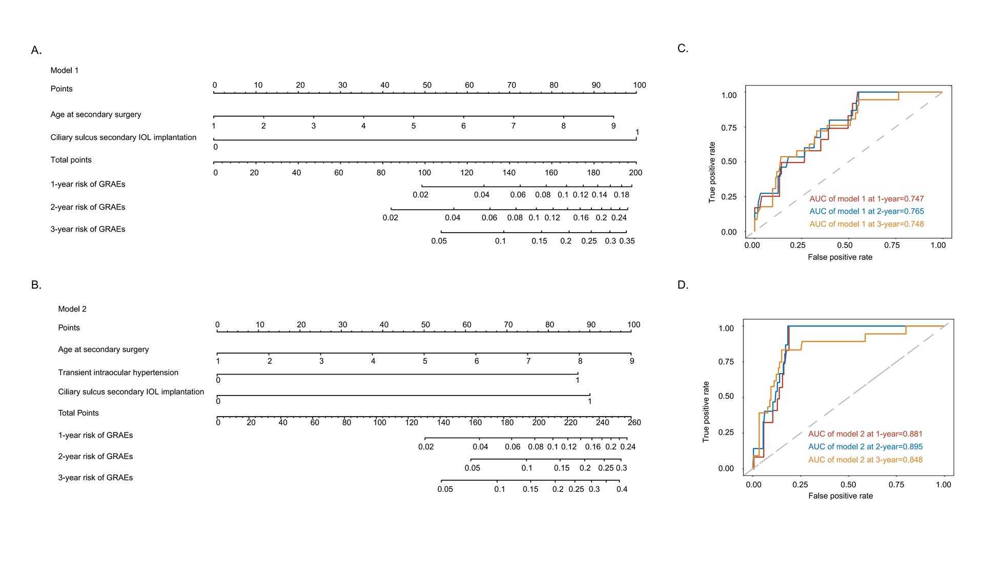
       考虑到一些临床环境可能缺乏患儿的随访信息,故使用不同的预测考虑到建多变量模型(表3)。多变量模型1包括两个不需要随访信息的预测因子,IOL睫状沟植入术(HR=11.36,9 5 % C I1.50~85.84,β=2.43, P=0.019)和行Ⅱ期IOL植入术时年龄较大(HR=1.33,95%CI 1.00~1.77;β= 0.29, P =0.047)。模型2包括一过性高眼压(HR=5.99,95%CI2.30~15.55,β=1.80, P<0.001),行Ⅱ期IOL植入术时年龄较大(HR=1.29,95%CI 0.97~1.73,β=0.26, P=0.085)和IOL睫状沟植入术(HR=6.37,95%CI 0.81~50.16,β= 1.85, P = 0.078)。基于此结果,本研究也构建了两个列线图,以提供Cox回归的可视化图形说明,见图1A、B。
       模型1为在随访1、2、3年时得到的AUC值,分别为0.747 (95% CI: 0.776~0.935)、0.765 (95% CI: 0.804~0.936)和0.748 (95% CI: 0.736~0.918),模型2的AUC值分别为0.881 (95% CI: 0.836~0.926)、0.895 (95%CI: 0.852~0.938)和0.848 (95% CI: 0.752~0.945)。见图1C、D。
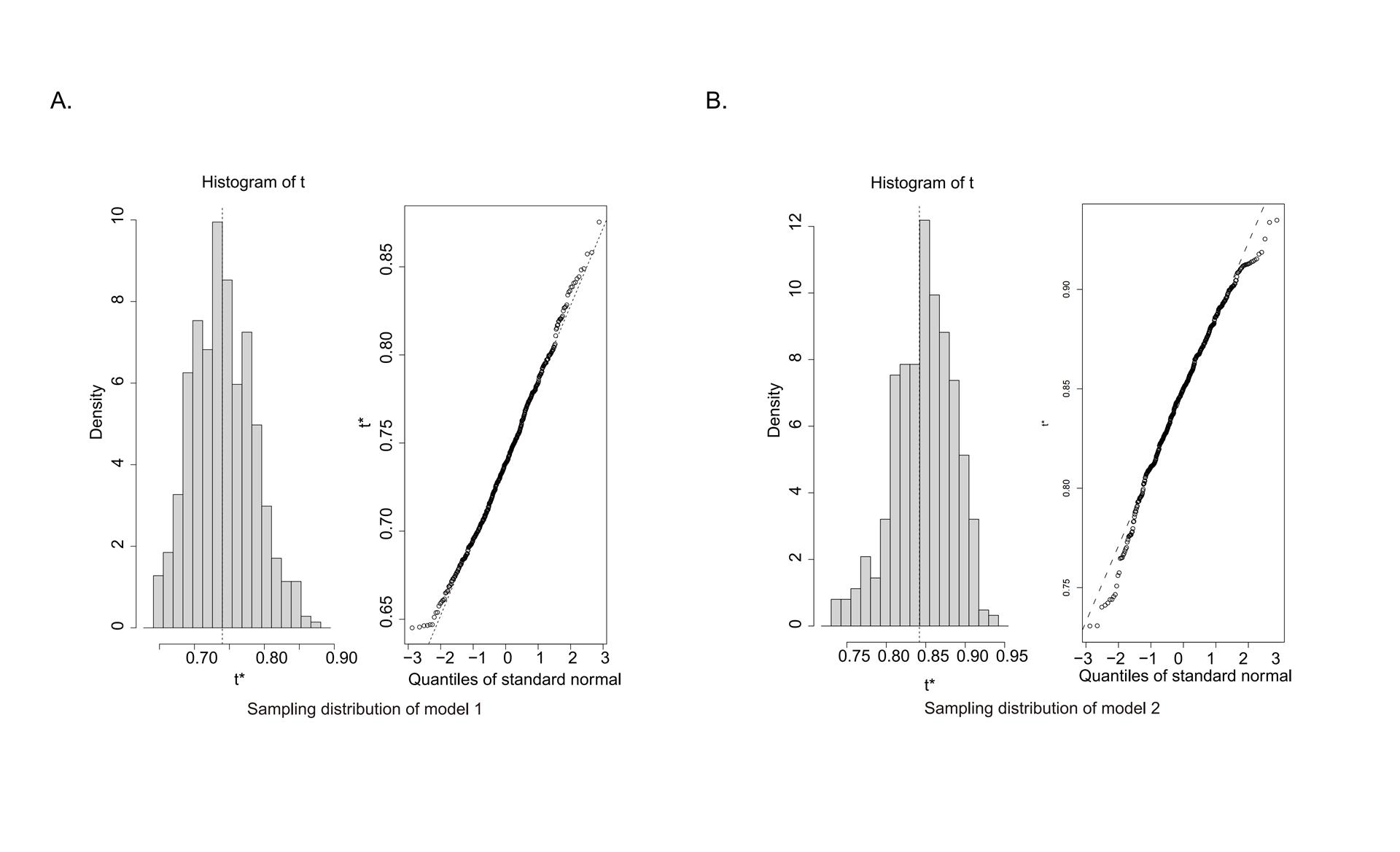
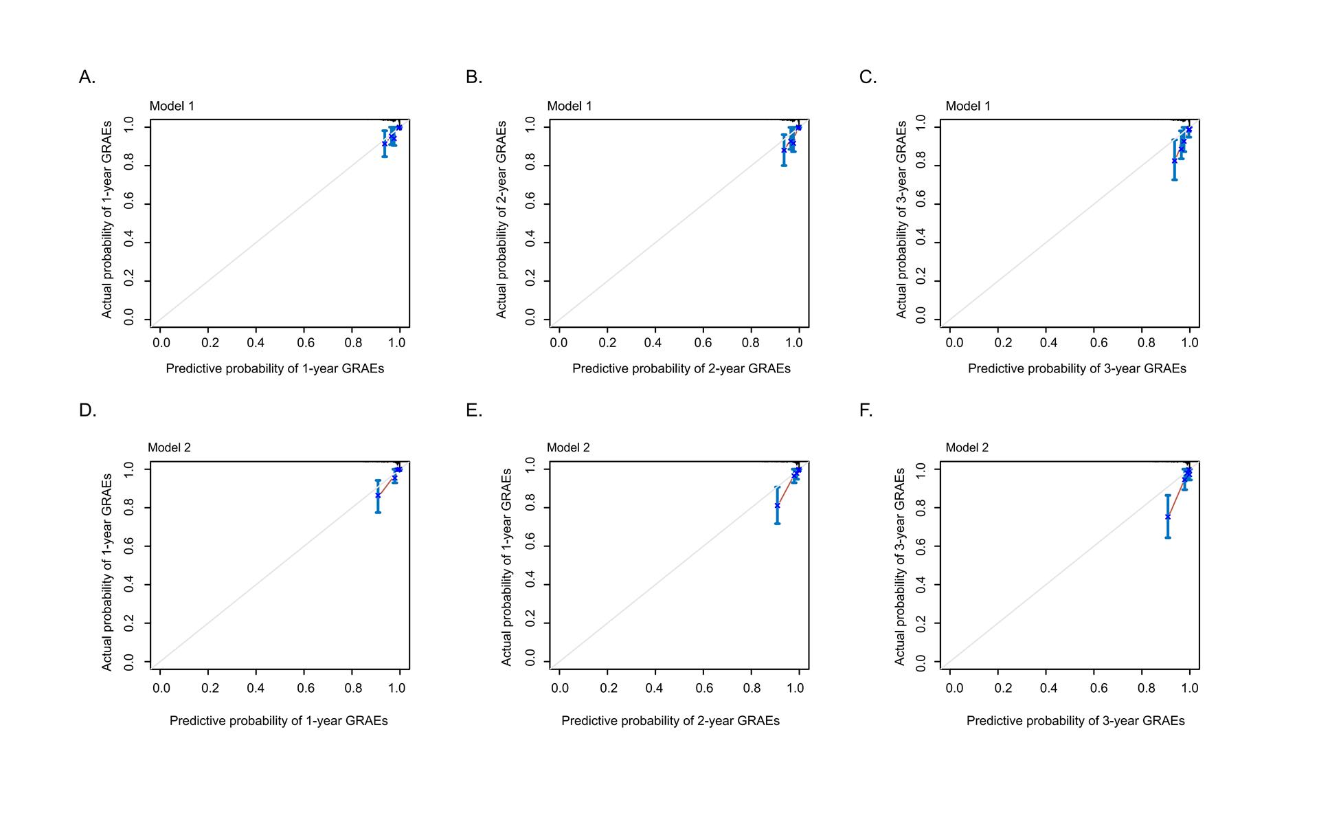
        采用Bootstrapping算法进行内部验证,模型1和模型2的Harrel's c指数分别为0.74 (95% CI: 0.65~0.82)和0.84 (95% CI: 0.77~0.91)(补充图1A、B)。校正图显示,对于GRAEs在术后第1、2、3年的预测,预测概率与观测发病率接近(补充图2A~F)
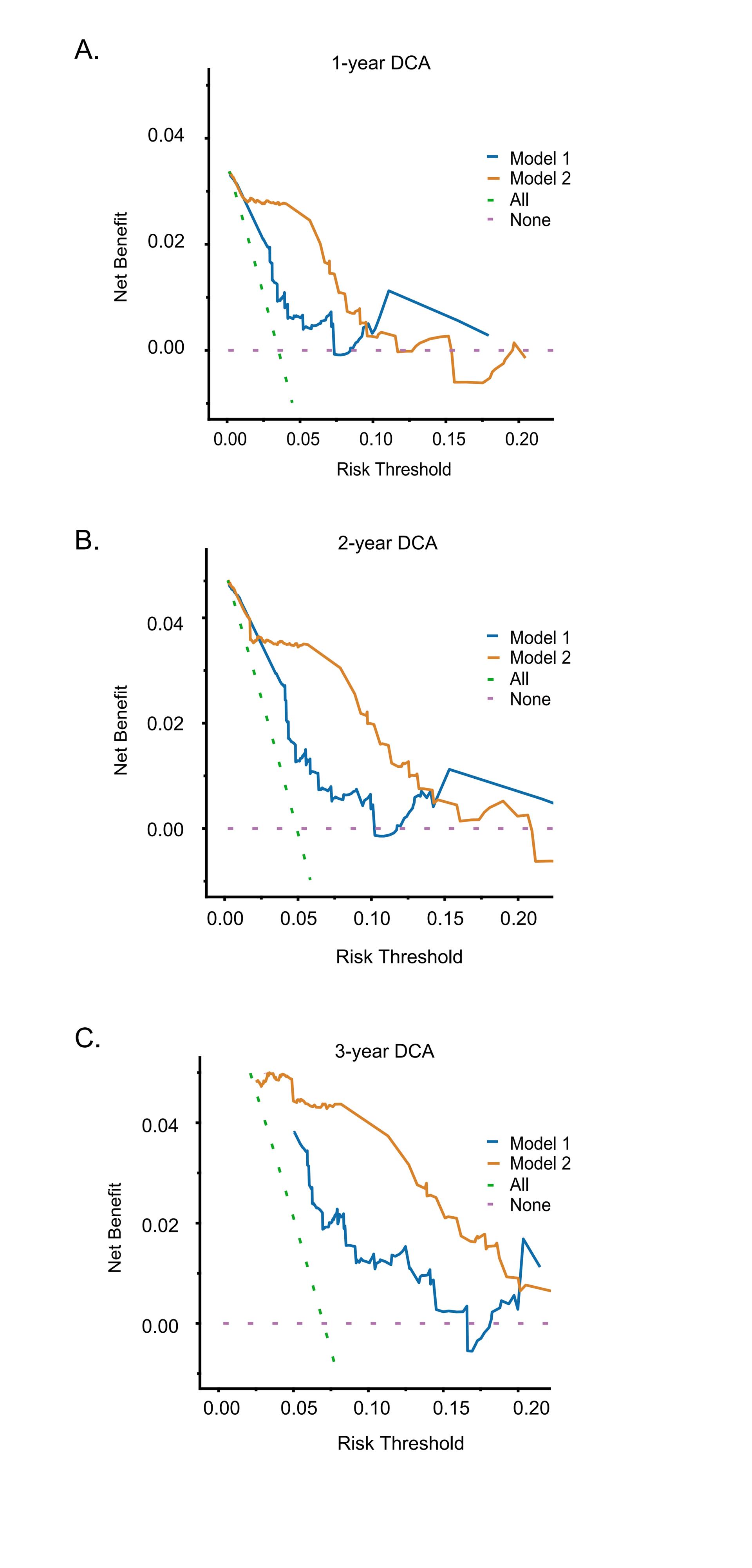
        在确定模型具有良好的辨别性和一致性后,进一步评价模型的临床应用价值。DCA显示,在较低的阈值下,模型2在随访第1、2和3年GRAEs预测方面优于模型1,并且这种建模方法使净临床效益最大化(补充图3A-C)
图1 模型1和模型2的列线图和基于时间依赖的ROC曲线
Figure 1 Nomograms and time-dependent receiver operating characteristic (ROC) curves of model 1 and model 2
(A)和(B)分别显示了模型1和模型2基于时间依赖的Cox风险比例分析的两个列线图。每一项参数都分别列出,参照顶部分数栏进行该参数分数的匹配。将每个变量的得分相加,生成总分,并在总分数线得分处向下画一条竖线,分别得到术后1、2、3年的GRAEs的对应概率;(C)和(D)分别显示了模型1和模型2在术后1、2、3年的ROC曲线,在随访的不同时间点显示AUC的值。AUC为曲线下面积;GRAEs为青光眼相关不良事件;IOL为人工晶状体。
Illustration of two nomograms based on time-dependent Cox hazard proportional analyses is shown in (A) and (B). The scores are listedseparately, and matching points are allocated to each other at the top. Scores from each variable are added to generate total points, and a verticalline is drawn from the total points to obtain the corresponding probability for GRAEs at postoperative 1, 2 and 3 years, respectively. ROC curvesof (C) model 1 and (D) model 2 at postoperative 1, 2 and 3 years are demonstrated. Values of AUC are displayed at different timepoints offollow-up visits. AUC, area under the curve; GRAEs, glaucoma-related adverse events; IOL, intraocular lens.
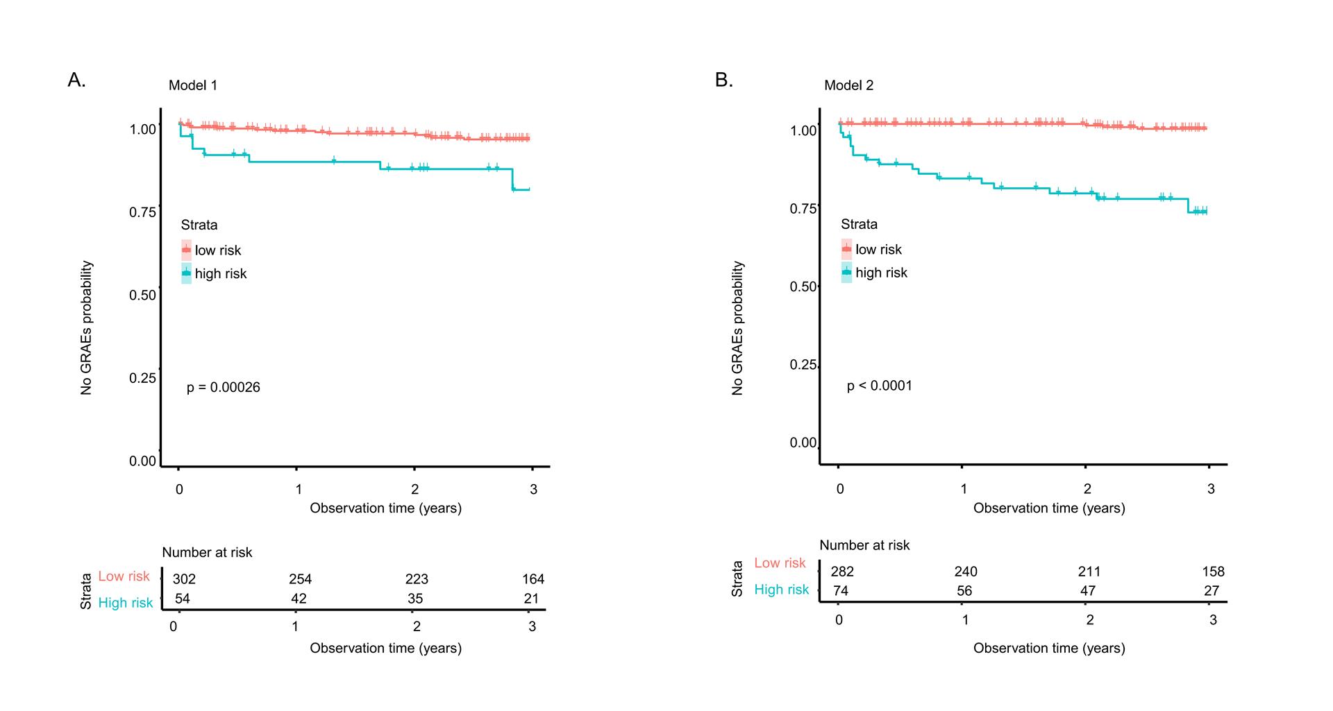
图2 Kaplan-Meier曲线显示了术后1年、2年和3年随访时(A)模型1和(B)模型2中参与者的分层情况。横轴代表术后时间,纵轴代表无青光眼相关不良事件(GRAEs)的概率。P值小于0.050被认为具有统计学意义。
Figure 2 Kaplan-Meier curves demonstrate the stratification of participants in (A) model 1 and (B) model 2 at postoperative 1-year,2-year and 3-year follow-up visits. The x-axis represents postoperative time and the y-axis represents probability without GRAEs. P values below 0.050 are regarded as statistically significant.

2.4 确定GRAEs风险分层,并建立儿童眼的在线预测系统

        Ⅱ期IOL植入术后的第1、2、3年时,模型1的截断值分别为0.073、0.100和0.144;模型2分别为0.076、0.105和0.148。图2A、B分别为两种模型中高危组和低危组在随访1、2、3年时的累积非GRAEs概率比较。
        按上述临界值分层时,高危组与低危组无GRAEs发生的概率比较差异均有统计学意义(均P<0.05)。为了使更多医生更加便捷地使用本模型,本研究还开发了在线的预测系统,免费提供给研究人员使用https://ccgrae.gzzoc.com:1002/。

3 讨论

        在这项对356只儿童眼进行的为期3年的队列研究中,我们证明了儿童Ⅱ期IOL植入术后GRAEs发生的三个预测因素。基于这些手术和术后预测因素的不同组合,构建了两个灵敏度和准确率较高的列线图来预测发生GRAEs的风险并应用于识别高危人群。
        本研究确定术后一过性高眼压是GRAEs的危险因素。接受白内障手术的儿童通常接受局部糖皮质激素(激素)治疗,这使他们容易发生短暂性IOP[22, 31]。然而,对激素性IOP的高易感性,或IOL植入术后1年内早期IOP波动与GRAEs发病之间的相关性值得进一步研究。此外,本研究结果表明,行 Ⅱ期IOL植入术时患儿的年龄越大,发生GRAEs的风险越高,换言之,行 Ⅱ期IOL植入术时患儿年龄越小则发生GRAEs的风险越低。这与其他一些研究结果形成对比。一种可能的推测是,在我们的队列中,年龄较小便接受Ⅱ期IOL植入术的患儿的眼能较早满足IOL植入术的严格条件,这表明他们可能比相对较大年龄时接受Ⅱ期IOL植入术的眼更早达到解剖成熟状态。这些结果和机制值得在不同的队列和更长的随访中进行进一步研究。
       此外,囊袋内IOL植入术已被证实可以预防GRAEs的发生,这与我们之前的队列研究结果相似[32]。睫状沟植入的IOL可能与虹膜直接接触,从而导致葡萄膜炎或前房角破坏,并伴有眼压升高[33]。先前的研究报道了接受Ⅱ期IOL植入术的眼发生GRAEs的一些危险因素,如眼轴较短[1, 11]、角膜直径较小[2]、前房较浅、中央角膜厚度较厚等[9]。然而,术前眼轴 (HR=0.98, 95%CI: 0.76~1.28, P=0.900)、前房深度(HR=0.708, 95%CI: 0.08~6.10, P=0.753)、中央角膜厚度(HR=1.00, 95%CI: 1.00 ~1.01,P=0.294)和前房容积(HR0.99, 95%CI: 0.98 ~1.00, P=0.129)在本队列中均无统计学意义。
        由于列线图可以提供简单而有效的预测风险说明,本研究开发了两个列线图,以图形展示随访1、2、3年期间的GRAEs预测风险。两种模型在不同随访期的AUC值均超过0.7,具有较强的预测能力。
        由于这两种预测模型所需的随访信息不同,因此可适用于术前、术后不同的临床环境。模型1仅包含手术参数(IOL植入位置和行IOL植入术时的年龄),在缺乏随访信息时可用于GRAEs预测,而在可获得一过性高眼压信息时,模型2可以更准确、灵敏地预测GRAEs的发生风险。通过在网站上输入不同模型所需的预测因子,可以方便地获得预测的GRAEs发生概率和风险分层。根据列线图所计算的风险结果进行分层,将受试者分为GRAEs发生的低危组和高危组,便于眼科医生对高危组进行更密切的观察或及时干预。
        本研究样本量较大,实验设计严格,随访方案规范,提出了儿童无晶状体眼GRAEs的预测模型。然而,在本研究中也存在不容忽视的一些局限性。本研究人群来自中国的一家三级甲等眼科中心,随访时间较短。因此,当将结果外推到其他种族背景的患儿时,应对所得的结果保持谨慎,并在未来的研究中需要更长的随访时间和外部验证。此外,小角膜、需要干预的视轴浑浊和需要干预的IOL脱位没有被确定为预测因素,可能是因为在我们的队列中这些情况的发病率很低。值得注意的是,由于8眼合并小角膜,另外210眼残留晶状体囊的量不足,而无法进行Ⅱ期IOL囊袋内植入。在我们的研究中,8只合并有小角膜的眼都没有发生GRAEs。先前的研究也报道过与GRAEs相关的其他因素,如永存胚胎血管[11]、先天性风疹综合征[8]、后囊膜状态和额外的眼内手术,这些因素也未被纳入本研究中[34]。本研究中后囊膜的状态是相同的,因为所有的患儿在白内障摘除过程中进行了后囊膜切除术和前段玻璃体切割术。此外,Ⅱ期IOL植入术的年龄和IOL植入的位置对GRAEs的预测作用较弱,这可能是因为该二者为其他眼部特征的相关因素而非致病原因。再者,由于排除了既往存在GRAEs的患儿,GRAEs的发生率可能被低估。同时,因为行Ⅱ期IOL植入术时患儿的平均年龄足够大,所以本队列的GRAEs风险可能有所降低。最后,值得注意的是,IOL植入囊袋或睫状沟的随机化也需在未来的研究中考虑。
        总之,基于迄今为止最大的接受Ⅱ期IOL植入术的儿童无晶状体眼患儿队列,我们提出了两种预测模型来对GRAEs风险进行个性化估计。研究建立的列线图模型可基于有或无Ⅱ期IOL植入术后的信息进行预测,以便眼科医生对这些患儿进行恰当的分层和管理,改善患儿的预后。

利益冲突

所有作者均声明不存在利益冲突。

开放获取声明

本文适用于知识共享许可协议 (Creative Commons),允许第三方用户按照署名(BY)-非商业性使用(NC)-禁止演绎(ND)(CC BY-NC-ND)的方式共享,即允许第三方对本刊发表的文章进行复制、发行、展览、表演、放映、广播或通过信息网络向公众传播,但在这些过程中必须保留作者署名、仅限于非商业性目的、不得进行演绎创作。详情请访问:https://creativecommons.org/licenses/by-nc-nd/4.0/。

版权声明

本文已获得BMJ出版集团的许可,对原文的中文翻译和正式发表。《眼科学报》对本文的中文翻译负有全部责任,包括译文中的错误、遗漏和其他不足之处。


补充图1
Supplementary fig.1
(A) 模型 1 和 (B) 模型 2 的一致性指数(t*)分布直方图和标准正态分布。
Histogram of the distribution of Harrel's C-statistic (t*) and standard normal distribution of Harrel's C-statistic of (A) model 1 and (B) model 2.


补充图2
Supplementary Fig.2
模型 1 在随访1 年(A)、2 年(B)和3 年(C)时的校准曲线。模型 2 在随访1 年(D)、2 年(E)和3 年(F)时的校准曲线。在校准曲线上,X 轴是术后不同时间点 GRAE 的预测概率,Y 轴是 GRAE 的实际概率。对角线代表理想预测,红色实线代表列线图的性能。与对角线越接近,表示预测效果越好。
Calibration curves of nomogram for model 1 at (A) 1-year, (B) 2-year and (C) 3-year follow-ups. Calibration curves of nomogram for model 2 at (D) 1-year, (E) 2-year and (F) 3-year follow-ups. On the calibration curve, the x-axis is the predictive probability for GRAEs at different postoperative timepoints, and the y-axis is the actual probability for GRAEs. The diagonal line represents the ideal prediction, and the red solid line represents the performance of nomogram. A closer fit to the diagonal line indicates a better prediction.
补充图3
Supplementary Fig.3
模型 1 和模型 2 在随访1 年(A)、2 年(B)和3 年(C)时的 DCA 曲线。DCA 曲线的横轴表示临界概率,纵轴表示净收益。斜虚线表示 "全部治疗 "的情况,横虚线表示 "未治疗 "的情况。在阈值概率相同的情况下,净获益越大,说明患儿通过使用预测模型获益越多。
DCA curves of model 1 and 2 at (A) 1-year, (B) 2-year and (C) 3-year follow-ups. The DCA curve abscissa shows the threshold probability, the ordinate is the net benefit. The slash dashed line represents the “all treated” situation, and the horizontal dashed line represents the “none treated” situation. Under the same threshold probability, the larger net benefit suggests that patients can benefit more by using the predictive model.
1、Liu Z, Lin H, Jin G, et al. In-the-bag versus ciliary sulcus secondary intraocular lens implantation for pediatric aphakia: a prospective comparative study[ J]. Am J Ophthalmol, 2022, 236: 183-192. DOI: 10.1016/j.ajo.2021.10.006.Liu Z, Lin H, Jin G, et al. In-the-bag versus ciliary sulcus secondary intraocular lens implantation for pediatric aphakia: a prospective comparative study[ J]. Am J Ophthalmol, 2022, 236: 183-192. DOI: 10.1016/j.ajo.2021.10.006.
2、Freedman SF, Lynn MJ, Beck AD, et al. Glaucoma-related adverse events in the first 5 years after unilateral cataract removal in the infant aphakia treatment study[ J]. JAMA Ophthalmol, 2015, 133(8): 907-914. DOI: 10.1001/jamaophthalmol.2015.1329.Freedman SF, Lynn MJ, Beck AD, et al. Glaucoma-related adverse events in the first 5 years after unilateral cataract removal in the infant aphakia treatment study[ J]. JAMA Ophthalmol, 2015, 133(8): 907-914. DOI: 10.1001/jamaophthalmol.2015.1329.
3、Papadopoulos M, Cable N, Rahi J, et al. The British infantile and childhood glaucoma (BIG) eye study[ J]. Invest Ophthalmol Vis Sci, 2007, 48(9): 4100-4106. DOI: 10.1167/iovs.06-1350.Papadopoulos M, Cable N, Rahi J, et al. The British infantile and childhood glaucoma (BIG) eye study[ J]. Invest Ophthalmol Vis Sci, 2007, 48(9): 4100-4106. DOI: 10.1167/iovs.06-1350.
4、Hoguet A, Grajewski A, Hodapp E, et al. A retrospective survey of childhood glaucoma prevalence according to Childhood Glaucoma Research Network classification[ J]. Indian J Ophthalmol, 2016, 64(2): 118-123. DOI: 10.4103/0301-4738.179716.Hoguet A, Grajewski A, Hodapp E, et al. A retrospective survey of childhood glaucoma prevalence according to Childhood Glaucoma Research Network classification[ J]. Indian J Ophthalmol, 2016, 64(2): 118-123. DOI: 10.4103/0301-4738.179716.
5、Beck A, Coleman T, Freedman S. Chapter 1: Definition, Classification, Differential Diagnosis[ J]. 2013: 3–10.Beck A, Coleman T, Freedman S. Chapter 1: Definition, Classification, Differential Diagnosis[ J]. 2013: 3–10.
6、Daniel MC, Dubis AM, Theodorou M, et al. Childhood lensectomy is associated with static and dynamic reduction in schlemm canal size: a biomechanical hypothesis of glaucoma after lensectomy[ J]. Ophthalmology, 2019, 126(2): 233-241. DOI: 10.1016/j.ophtha.2018.08.031.Daniel MC, Dubis AM, Theodorou M, et al. Childhood lensectomy is associated with static and dynamic reduction in schlemm canal size: a biomechanical hypothesis of glaucoma after lensectomy[ J]. Ophthalmology, 2019, 126(2): 233-241. DOI: 10.1016/j.ophtha.2018.08.031.
7、Freedman SF, Beck AD, Nizam A, et al. Glaucoma-related adverse events at 10 years in the infant aphakia treatment study: a secondary analysis of a randomized clinical trial[ J]. JAMA Ophthalmol, 2021, 139(2): 165-173. DOI: 10.1001/jamaophthalmol.2020.5664.Freedman SF, Beck AD, Nizam A, et al. Glaucoma-related adverse events at 10 years in the infant aphakia treatment study: a secondary analysis of a randomized clinical trial[ J]. JAMA Ophthalmol, 2021, 139(2): 165-173. DOI: 10.1001/jamaophthalmol.2020.5664.
8、Shah SK, Praveen MR, Vasavada AR, et al. Long-term longitudinal assessment of postoperative outcomes after congenital cataract surgery in children with congenital rubella syndrome[ J]. J Cataract Refract Surg, 2014, 40(12): 2091-2098. DOI: 10.1016/j.jcrs.2014.04.028.Shah SK, Praveen MR, Vasavada AR, et al. Long-term longitudinal assessment of postoperative outcomes after congenital cataract surgery in children with congenital rubella syndrome[ J]. J Cataract Refract Surg, 2014, 40(12): 2091-2098. DOI: 10.1016/j.jcrs.2014.04.028.
9、Wang J, Chen J, Chen W, et al. Incidence of and risk factors for suspected glaucoma and glaucoma after congenital and infantile cataract surgery: a longitudinal study in China[ J]. J Glaucoma, 2020, 29(1): 46-52. DOI: 10.1097/IJG.0000000000001398.Wang J, Chen J, Chen W, et al. Incidence of and risk factors for suspected glaucoma and glaucoma after congenital and infantile cataract surgery: a longitudinal study in China[ J]. J Glaucoma, 2020, 29(1): 46-52. DOI: 10.1097/IJG.0000000000001398.
10、Chak M, Rahi JS, Group BCCI. Incidence of and factors associated with glaucoma after surgery for congenital cataract: findings from the British Congenital Cataract Study[ J]. Ophthalmology, 2008, 115(6): 1013-1018.e2. DOI: 10.1016/j.ophtha.2007.09.002.Chak M, Rahi JS, Group BCCI. Incidence of and factors associated with glaucoma after surgery for congenital cataract: findings from the British Congenital Cataract Study[ J]. Ophthalmology, 2008, 115(6): 1013-1018.e2. DOI: 10.1016/j.ophtha.2007.09.002.
11、Solebo AL, Rahi JS, Group BCCI. Glaucoma following cataract surgery in the first 2 years of life: frequency, risk factors and outcomes from IoLunder2[ J]. Br J Ophthalmol, 2020, 104(7): 967-973. DOI: 10.1136/bjophthalmol-2019-314804.Solebo AL, Rahi JS, Group BCCI. Glaucoma following cataract surgery in the first 2 years of life: frequency, risk factors and outcomes from IoLunder2[ J]. Br J Ophthalmol, 2020, 104(7): 967-973. DOI: 10.1136/bjophthalmol-2019-314804.
12、Bothun ED, Wilson ME, Vanderveen DK, et al. Outcomes of bilateral cataracts removed in infants 1 to 7 months of age using the toddler aphakia and pseudophakia treatment study registry[ J]. Ophthalmology, 2020, 127(4): 501-510. DOI: 10.1016/j.ophtha.2019.10.039.Bothun ED, Wilson ME, Vanderveen DK, et al. Outcomes of bilateral cataracts removed in infants 1 to 7 months of age using the toddler aphakia and pseudophakia treatment study registry[ J]. Ophthalmology, 2020, 127(4): 501-510. DOI: 10.1016/j.ophtha.2019.10.039.
13、Leske MC, Wu SY, Hennis A, et al. Risk factors for incident open-angle glaucoma: the Barbados Eye Studies[ J]. Ophthalmology, 2008, 115(1): 85-93. DOI: 10.1016/j.ophtha.2007.03.017.Leske MC, Wu SY, Hennis A, et al. Risk factors for incident open-angle glaucoma: the Barbados Eye Studies[ J]. Ophthalmology, 2008, 115(1): 85-93. DOI: 10.1016/j.ophtha.2007.03.017.
14、Bothun ED, Wilson ME, Traboulsi EI, et al. Anderson JS, Lambert SR; Toddler Aphakia and Pseudophakia Study Group (TAPS). Outcomes of Unilateral Cataracts in Infants and Toddlers 7 to 24 Months of Age: Toddler Aphakia and Pseudophakia Study (TAPS) [ J]. Ophthalmology. 2019 Aug;126(8):1189-1195. doi: 10.1016/j.ophtha.2019.03.011.Bothun ED, Wilson ME, Traboulsi EI, et al. Anderson JS, Lambert SR; Toddler Aphakia and Pseudophakia Study Group (TAPS). Outcomes of Unilateral Cataracts in Infants and Toddlers 7 to 24 Months of Age: Toddler Aphakia and Pseudophakia Study (TAPS) [ J]. Ophthalmology. 2019 Aug;126(8):1189-1195. doi: 10.1016/j.ophtha.2019.03.011.
15、Iasonos A, Schrag D, Raj GV, et al. How to build and interpret a nomogram for cancer prognosis[ J]. J Clin Oncol, 2008, 26(8): 1364-1370. DOI: 10.1200/JCO.2007.12.9791.Iasonos A, Schrag D, Raj GV, et al. How to build and interpret a nomogram for cancer prognosis[ J]. J Clin Oncol, 2008, 26(8): 1364-1370. DOI: 10.1200/JCO.2007.12.9791.
16、Wang R, Dai W, Gong J, et al. Development of a novel combined nomogram model integrating deep learning-pathomics, radiomics and immunoscore to predict postoperative outcome of colorectal cancer lung metastasis patients[ J]. J Hematol Oncol, 2022, 15(1): 11. DOI: 10.1186/s13045-022-01225-3.Wang R, Dai W, Gong J, et al. Development of a novel combined nomogram model integrating deep learning-pathomics, radiomics and immunoscore to predict postoperative outcome of colorectal cancer lung metastasis patients[ J]. J Hematol Oncol, 2022, 15(1): 11. DOI: 10.1186/s13045-022-01225-3.
17、Li Q, Jiang T, Zhang C, et al. A nomogram based on clinical information, conventional ultrasound and radiomics improves prediction of malignant parotid gland lesions[ J]. Cancer Lett, 2022, 527: 107-114. DOI: 10.1016/j.canlet.2021.12.015.Li Q, Jiang T, Zhang C, et al. A nomogram based on clinical information, conventional ultrasound and radiomics improves prediction of malignant parotid gland lesions[ J]. Cancer Lett, 2022, 527: 107-114. DOI: 10.1016/j.canlet.2021.12.015.
18、Zhang J, Han X, Zhang M, et al. Predicting the risk of clinically significant intraocular lens tilt and decentration in vitrectomized eyes[ J]. J Cataract Refract Surg, 2022, 48(11): 1318-1324. DOI: 10.1097/j.jcrs.0000000000000997.Zhang J, Han X, Zhang M, et al. Predicting the risk of clinically significant intraocular lens tilt and decentration in vitrectomized eyes[ J]. J Cataract Refract Surg, 2022, 48(11): 1318-1324. DOI: 10.1097/j.jcrs.0000000000000997.
19、White RR, Kattan MW, Haney JC, et al. Evaluation of preoperative therapy for pancreatic cancer using a prognostic nomogram[ J]. Ann Surg Oncol, 2006, 13(11): 1485-1492. DOI: 10.1245/s10434-006-9104-y.White RR, Kattan MW, Haney JC, et al. Evaluation of preoperative therapy for pancreatic cancer using a prognostic nomogram[ J]. Ann Surg Oncol, 2006, 13(11): 1485-1492. DOI: 10.1245/s10434-006-9104-y.
20、Roach M 3rd, Weinberg V, Nash M, et al. Defining high risk prostate cancer with risk groups and nomograms: implications for designing clinical trials[ J]. J Urol, 2006, 176(6 Pt 2): S16-S20. DOI: 10.1016/j.juro.2006.06.081.Roach M 3rd, Weinberg V, Nash M, et al. Defining high risk prostate cancer with risk groups and nomograms: implications for designing clinical trials[ J]. J Urol, 2006, 176(6 Pt 2): S16-S20. DOI: 10.1016/j.juro.2006.06.081.
21、Lin H, Chen W, Luo L, et al. Effectiveness of a short message reminder in increasing compliance with pediatric cataract treatment: a randomized trial[ J]. Ophthalmology, 2012, 119(12): 2463-2470. DOI: 10.1016/j.ophtha.2012.06.046.Lin H, Chen W, Luo L, et al. Effectiveness of a short message reminder in increasing compliance with pediatric cataract treatment: a randomized trial[ J]. Ophthalmology, 2012, 119(12): 2463-2470. DOI: 10.1016/j.ophtha.2012.06.046.
22、Lin H, Chen W, Luo L, et al. Ocular hypertension after pediatric cataract surgery: baseline characteristics and first-year report[ J]. PLoS One, 2013, 8(7): e69867. DOI: 10.1371/journal.pone.0069867.Lin H, Chen W, Luo L, et al. Ocular hypertension after pediatric cataract surgery: baseline characteristics and first-year report[ J]. PLoS One, 2013, 8(7): e69867. DOI: 10.1371/journal.pone.0069867.
23、Beck AD, Freedman SF, Lynn MJ, et al. Glaucoma-related adverseevents in the Infant Aphakia Treatment Study: 1-year results[ J]. Arch Ophthalmol. 2012 Mar;130(3):300-5. doi: 10.1001/archophthalmol.2011.347.Beck AD, Freedman SF, Lynn MJ, et al. Glaucoma-related adverseevents in the Infant Aphakia Treatment Study: 1-year results[ J]. Arch Ophthalmol. 2012 Mar;130(3):300-5. doi: 10.1001/archophthalmol.2011.347.
24、Chen X, Gu X, Wang W, et al. Characteristics and factors associated with intraocular lens tilt and decentration after cataract surgery[ J]. J Cataract Refract Surg, 2020, 46(8): 1126-1131. DOI: 10.1097/j.jcrs.0000000000000219.Chen X, Gu X, Wang W, et al. Characteristics and factors associated with intraocular lens tilt and decentration after cataract surgery[ J]. J Cataract Refract Surg, 2020, 46(8): 1126-1131. DOI: 10.1097/j.jcrs.0000000000000219.
25、Liu Z, Zou Y, Yu Y, et al. Accuracy of Intraocular Lens Power Calculation in Pediatric Secondary Implantation: In-the-Bag Versus Sulcus Placement[ J]. Am J Ophthalmol. 2023 May;249:137-143. doi: 10.1016/j.ajo.2022.12.028.Liu Z, Zou Y, Yu Y, et al. Accuracy of Intraocular Lens Power Calculation in Pediatric Secondary Implantation: In-the-Bag Versus Sulcus Placement[ J]. Am J Ophthalmol. 2023 May;249:137-143. doi: 10.1016/j.ajo.2022.12.028.
26、Royston P. Multiple imputation of missing values[ J]. Stata J, 2004, 4: 227-241. DOI: 10.1177/1536867X0400400301.Royston P. Multiple imputation of missing values[ J]. Stata J, 2004, 4: 227-241. DOI: 10.1177/1536867X0400400301.
27、Huang YQ, Liang CH, He L, et al. Development and validation of a radiomics nomogram for preoperative prediction of lymph node metastasis in colorectal cancer[ J]. J Clin Oncol, 2016, 34(18): 2157-2164. DOI: 10.1200/JCO.2015.65.9128.Huang YQ, Liang CH, He L, et al. Development and validation of a radiomics nomogram for preoperative prediction of lymph node metastasis in colorectal cancer[ J]. J Clin Oncol, 2016, 34(18): 2157-2164. DOI: 10.1200/JCO.2015.65.9128.
28、Cadrin-Tourigny J, Bosman LP, Nozza A, et al. A new prediction model for ventricular arrhythmias in arrhythmogenic right ventricular cardiomyopathy[ J]. Eur Heart J, 2022, 43(32): e1-e9. DOI: 10.1093/eurheartj/ehac180.Cadrin-Tourigny J, Bosman LP, Nozza A, et al. A new prediction model for ventricular arrhythmias in arrhythmogenic right ventricular cardiomyopathy[ J]. Eur Heart J, 2022, 43(32): e1-e9. DOI: 10.1093/eurheartj/ehac180.
29、Camp RL, Dolled-Filhart M, Rimm DL. X-tile: a new bio-informatics tool for biomarker assessment and outcome-based cut-point optimization[ J]. Clin Cancer Res, 2004, 10(21): 7252-7259. DOI: 10.1158/1078-0432.CCR-04-0713.Camp RL, Dolled-Filhart M, Rimm DL. X-tile: a new bio-informatics tool for biomarker assessment and outcome-based cut-point optimization[ J]. Clin Cancer Res, 2004, 10(21): 7252-7259. DOI: 10.1158/1078-0432.CCR-04-0713.
30、Zhang JX, Song W, Chen ZH, et al. Prognostic and predictive value of a microRNA signature in stage II colon cancer: a microRNA expression analysis[ J]. Lancet Oncol, 2013, 14(13): 1295-1306. DOI: 10.1016/S1470-2045(13)70491-1.Zhang JX, Song W, Chen ZH, et al. Prognostic and predictive value of a microRNA signature in stage II colon cancer: a microRNA expression analysis[ J]. Lancet Oncol, 2013, 14(13): 1295-1306. DOI: 10.1016/S1470-2045(13)70491-1.
31、Zhang Z, Fu Y, Wang J, et al. Glaucoma and risk factors three years after congenital cataract surgery[ J]. BMC Ophthalmol, 2022, 22(1): 118. DOI: 10.1186/s12886-022-02343-9.Zhang Z, Fu Y, Wang J, et al. Glaucoma and risk factors three years after congenital cataract surgery[ J]. BMC Ophthalmol, 2022, 22(1): 118. DOI: 10.1186/s12886-022-02343-9.
32、Shenoy BH, Mittal V, Gupta A, et al. Complications and visual outcomes after secondary intraocular lens implantation in children[ J]. Am J Ophthalmol, 2015, 159(4): 720-726. DOI: 10.1016/j.ajo.2015.01.002.Shenoy BH, Mittal V, Gupta A, et al. Complications and visual outcomes after secondary intraocular lens implantation in children[ J]. Am J Ophthalmol, 2015, 159(4): 720-726. DOI: 10.1016/j.ajo.2015.01.002.
33、Wu X, Liu Z, Wang D, et al. Preoperative profile of inflammatory factors in aqueous humor correlates with postoperative inflammatory response in patients with congenital cataract[ J]. Mol Vis, 2018, 24: 414-424.Wu X, Liu Z, Wang D, et al. Preoperative profile of inflammatory factors in aqueous humor correlates with postoperative inflammatory response in patients with congenital cataract[ J]. Mol Vis, 2018, 24: 414-424.
34、Zhang Y, Song Y, Zhou Y, et al. A comprehensive review of pediatric glaucoma following cataract surgery and progress in treatment[ J]. Asia Pacific J Ophthalmol, 12(1):94-102.Zhang Y, Song Y, Zhou Y, et al. A comprehensive review of pediatric glaucoma following cataract surgery and progress in treatment[ J]. Asia Pacific J Ophthalmol, 12(1):94-102.
1、 广州市科技计划 (202201011815)。This work was supported by the Science and Technology Program of Guangzhou, China, (202201011815).()
上一篇
下一篇
其他期刊
  • 眼科学报

    主管:中华人民共和国教育部
    主办:中山大学
    承办:中山大学中山眼科中心
    主编:林浩添
    主管:中华人民共和国教育部
    主办:中山大学
    浏览
  • Eye Science

    主管:中华人民共和国教育部
    主办:中山大学
    承办:中山大学中山眼科中心
    主编:林浩添
    主管:中华人民共和国教育部
    主办:中山大学
    浏览
推荐阅读
出版者信息
目录