您的位置: 首页 > 2008年3月 第24卷 第1期 > 文字全文
2023年7月 第38卷 第7期11
目录

bFGF、EGF和NGF对人角膜内皮细胞生长调控的实验研究

A Study on Effect of bFGF、EGF and NGF on Growth of Cultured Human Corneal Endothelial Cells

来源期刊: 眼科学报 | 2008年3月 第24卷 第1期 9-12 发布时间: 收稿时间:2024/10/16 15:11:25 阅读量:115
作者:
关键词:
角膜内皮细胞生长因子调控
Coreal endothelial cell Growth factor Regulation of growth
DOI:
收稿时间:
 
修订日期:
 
接收日期:
 
目的:探讨碱性成纤维细胞生长因子(Basic fibroblast growthfacfor, bFCF),表皮细胞生长因子(Epidermal growth factor, EGF)和神经细胞生长因子(Nerve growth factor, NGF)对体外培养的人角膜内皮细胞的生长调控作用。
方法:将相同数量的人角膜内皮细胞接种于96孔板。加人浓度分别为0 ng/ml、1 ng/ml、3 ng/ml、10 ng/ml、30 ng/ml、100 ng/ml的 EGF、bFGF 和 NGF 进行培养。5 天后 MTT法用检测增殖情况。
结果:在0 ng/ml、1 ng/ml、3 ng/ml、10 ng/ml、30 ng/ml、100 ng/ml 浓度下 bFGF 组的平均 OD 值分别为: 0.224±0.045、0.239±0.040、0.262±0.0342、0.278±0.0319、0.281±0.0324、0.260±0.0310 EGF组的平均 0D 值分别为: 0.228±0.0304、0.245±0.0418、0.267±0.0454、0.275±0.0347、0.271±0.0449、0.250±0.0253。NGF 组的平均 OD 值分别为:0.216±0.0187、0.228±0.0226、0.231±0.0225、0.242±0.0279、0.245±0.0294、0.247±0.0349。
结论:bFGF在 30 ng/ml范围内对内皮细胞生长有促进作用,并具有剂量依赖性。高于100 ng/ml时促生长作用降低。EGF在10 ng/ml范围内对内皮细胞生长有促进作用,并具有剂量依赖性。高于30 ng/ml 时促生长作用降低。NGF本次实验剂量范围内对角膜内皮细胞生长无明显作用。
Purpose: To investigate effect of bFGF, EGF and NGF on growth of cultured humancorneal endothelial cells.
Methods: Cultured human corneal endothelial cells were seeded into individual wellsof 96-well tissue culture plate with the same culture media containing separately bFCF, EGF or NGF with a serial of concentrations of 0 ng/ml、1 ng/ml 、3 ng/ml、10 ng/ml 、30 ng/ml and 100 ng/ml and then cultured for 5 days. Then MTT method wasused to detect the growth of the cells.
Results: The averaged OD values of the cell wells containing bFCF with a serial of concentrations of 0 ng/ml、1 ng/ml、3 ng/ml、10 ng/ml、30 ng/ml and 100 ng/ml were 0.224±0.045, 0.239±0.040, 0.262±0.0342, 0.278±0.0319, 0.281±0.0324, 0.260±0.0310. The averaged OD values of EGF group and NGF group were separately 0.228±0.0304,0.245±0.0418, 0.267±0.0454, 0.275±0.0347, 0.271±0.0449, 0.250±0.0253 and 0.216±0.0187, 0.228±0.0226, 0.231±0.0225, 0.242±0.0279, 0.245±0.0294,0.247±0.0349.
Conclusion: bGFC can promote the growth of human corneal endothelial cells in adose dependent manner while with concentration lower than 30 ng/ml. bFGF withconcentration that is higher than 100ng/ml will weaken this effect. EGF can alsopromote proliferation of human corneal endothelial cells demonstrating a linear dosedependent effect when its concentration is lower than 10 ng/ml and this effect decreasedwhen its concentration was higher than 30 ng/ml, NGF showed no effect on the growthof human cornea endothelial cells in this study.
       体外培养扩增人角膜内皮细胞目前仍然是一个世界性的难题。大量的研究表明,肽类细胞生长因子具有促进细胞生长及调节细胞功能的作用。因此,我们对几种常见的细胞生长因子,即碱性成纤维细胞生长因子(Basicfbroblast growthfactor, bFGF)、表皮细胞生长因子(Epidermal growthfactor, ECF)和神经细胞生长因子(Nerve growthfactor, NGF)在对体外培养的人角膜内皮细胞生长调控中的作用进行了初步探讨,现报道如下。

材料与方法

一、材料

       本研究的内皮细胞来自于手术室提供的,保存于 Optisol保存液中的穿透性角膜移植术后的剩余角膜组织。选取的材料,供体年龄在 20~40 岁之间,保存时间少于7 d。将角膜组织块从保存瓶中取出,用 2 ml D-Hank' s液漂洗2次,放人装有 2 ml含1%胎牛血清DMEM/F12(1∶1)混合液的25 mm培养皿内,置于37℃,含5% C02的潮湿培养箱内孵育 24 h。之后,将其取出,在解剖显微镜下,用显微无齿镊将后弹力膜撕下,用2 ml D-Hank' s漂洗2次后放人装有1 ml上述相同培养液的24孔板内,加人1 ml 0.4% collagenase Ⅳ消化(CA)1 h。吸出,注人15 ml离心管内。再加人9 ml培养液,1000转/分离心5 min。弃去上清液,加入培养液1 ml用巴斯德管吹打5次,注人24孔板,置于37℃,含5%C02的潮湿培养箱内培养。3 d后细胞已全部贴壁,得到原代细胞。待细胞融合为单层细胞且面积达培养孔面积的 90%以上时,进行传代。将培养液吸出,加入1 ml PBS,轻微倾斜培养板数次。吸出PBS,加人0.5 ml 0.25%胰蛋白酶+0.02%EDTA,在37℃下消化,并在倒置相差显微镜观察。当有50%左右细胞脱离培养孔底部时加入0.5ml培养液中和,轻微吹打后将细胞悬液吸出注入15 ml离心管,1000转/分离心5 min。弃去上清液,加入培养液1 ml,用巴斯德管吹打5次后进行细胞计数。取0.04 ml细胞悬液滴人房室培养板,加人0.3 ml培养液培养至贴壁。其中1个房室种入角膜基质细胞,作为对照组。用抗S-100抗体进行免疫组化染色鉴定。角膜内皮细胞呈阳性反应,角膜基质细胞呈阴性反应。将剩余的细胞接种于25 cm2培养瓶中,再加人相同培养液 2.5 ml。置于37℃,含5% CO2的潮湿培养箱内培养。3 d后细胞已全部贴壁,得到二代细胞。隔天换液,至融合为单层的细胞面积达培养瓶面积的 70%~90%,供下一步实验使用。

二、方法

       将25 cm2培养瓶中培养液吸出,加入1 mlPBS,轻微倾斜培养板数次。吸出PBS,加入0.5 ml 0.25%胰蛋白酶+0.02%EDTA,在37℃下消化,并在倒置相差显微镜观察。当有50%左右细胞脱离培养孔底部时,加人0.5 ml培养液中和,轻微吹打后将细胞悬液吸出注人15 ml离心管,1000转/分离心5 min。弃去上清液,加人培养液1 ml,用巴斯德管吹打5次后进行细胞计数。在96孔板上每个孔内分别接种内皮细胞约 5x10个。每孔加DMEM/F12培养基(1:1)培养基200μ l,共72个孔。重复接种3块96 孔板。培养24  h。每4个孔一组,每组分别更换200 μ l的下述培养液:含bFGF 0 ng/ml、1 ng/ml、3 ng/ml、10 ng/ml、30 ng/ml、100 ng/ml的DMEM/FI2 培养基(1:1)培养基,含EGF 0 ng/ml、l ng/ml、3 ng/ml、10 ng/ml、30 ng/ml、100 ng/ml的DMEM/F12 培养基(1:1)培养基和含 NGF 0 ng/ml、1 ng/ml、3 ng/ml、10 ng/ml、30 ng/ml、100 ng/ml的DMEM/FI2 培养基(1:1)培养基。在4个未加细胞的空白孔内加人200 μ DMEM /F12培养基(1:1)培养基作为空白对照。每天换液。5 d后用 MTT法检测细胞增殖生长情况。每孔加入5 mg/ml MTT的溶液 20 ml,继续培养4 h。弃去培养液。每孔加入DMSO 200 ml 37℃下摇匀,10 min。选择波长 570 nm 检测光源,波长 630 nm为校正光源,以未加细胞的空白孔作为对照。用酶标仪进行检测。以 OD 值绘制生长曲线。

结果

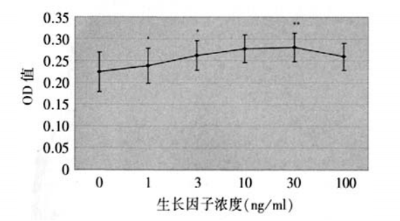
一、bFCF对角膜内皮细胞生长的影响(图1)

       在0 ng/ml、1 ng/ml、3 ng/ml、10 ng/ml、30 ng/ml、100 ng/ml 浓度下平均 OD 值分别为:0.224±0.045、0.239±0.040、0.262±0.0 342、0.278 ±0.0 319、0.281±0.0 324、0.260±0.0310 bFGF。表明 bFGF 在 30 ng/ml浓度以内对角膜内皮细胞生长具有促进作用,并有剂量依赖性。100 ng/ml浓度时促生长作用下降。
图1 bFCF对人角膜内皮细胞生长影响的生长曲线(*表示 P<0.01,**表示 P<0.05)

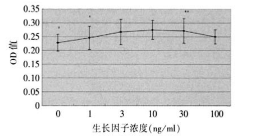
二、ECF 对角膜内皮细胞生长的影响(图2)

       在0 ng/ml、1 ng/ml、3 ng/ml 、10 ng/ml、30 ng/ml、100 ng/ml 浓度下平均 OD 值分别为:0.228±0.0 304、0.245±0.0 418、0.267±0.0 454、0.275±0.0 347、0.271±0.0449、0.250±0.0253。表明EGF 在 10 ng/ml 浓度以内对角膜内皮细胞生长具有促进作用,并有剂量依赖性。超过10 ng/ml 浓度则失去剂量依赖。100 ng/ml 浓度时促生长作用下降。

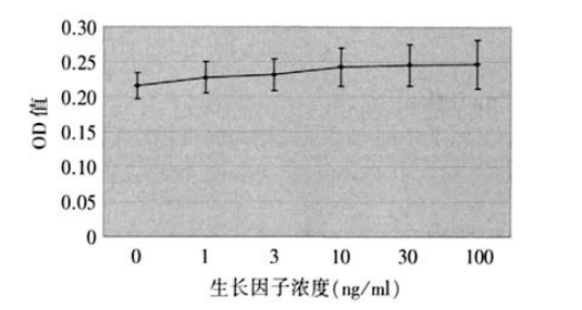
三、NGF对角膜内皮细胞生长的影响(图3)

       在0 ng/ml、1 ng/ml、3 ng/ml、10 ng/ml、30 ng/ml、100 ng/ml 浓度下平均 OD 值分别为:0.216±0.0187、0.228±0.0 226、0.231±0.0 225、0.242±0.0 279、0.245±0.0 294、0.247±0.0 349。表明NGF在本实验浓度范围内,NGF对角膜内皮细胞生长无明显促进作用。
          图2 ECF对人角膜内皮细胞生长影响的生长曲线(表示P<0.01,**表示P<0.05)      图3'NGF对人角膜内皮细胞生长影响的生长曲线(均P>0.05)

讨论

       在体外对人角膜内皮细胞进行培养、扩增,并种植于适当的载体,即体外构建组织工程角膜内皮植片,是克服供体材料来源紧缺的瓶颈的最直接和最有希望的途径。然而,体外构建组织工程角膜内皮植片的关键问题是怎样取得足够多的人角膜内皮细胞。
       在正常的生理条件下,人角膜内皮细胞不增殖或增殖速度低于因衰老而损失的速度[1]。以至于成年人的角膜内皮细胞密度以每年 0.3%~0.6%的速率下降。许多学者尝试在体外人工培养的条件下对成熟的人角膜内皮细胞进行扩增,但是未能取得理想的效果[2]。体外有效扩增人角膜内皮细胞仍是一个世界性的难题。
       细胞生长因子是一类具有调节多种生理过程的多功能调节蛋白,肽类生长因子具有促进或抑制细胞生长及调节细胞功能的作用。bFGF、ECF和NGF均为最常见的细胞生长因子[3]。文献表明,人角膜内皮细胞可表达bFGF、EGF的受体[4]。本课题组研究发现人角膜内皮细胞可表达bFGFEGF和NGF等生长因子及其受体的mRNA。Yu等[5]发现bFGF、EGF和NGF均对哺乳动物的角膜内皮细胞生长有促进作用。
       老年性白内障患者房水中的bFGF浓度为 0.4~0.8 ng/ml[6]。放免法测定新鲜人房水样本中的bFGF浓度为 0.48~1.44 ng/ml[7]。有关 bFGF 对角膜内皮细胞的促增殖作用已有较多报道。Johnstone等的实验显示:bFCF可以增加细胞的最终密度但是高于25 ng/ml浓度的FCF会使角膜内皮细胞的形态变成接近成纤维细胞的形态[8]
       文献报道在人房水中未能检测到 EGF[6],推测可能其浓度在检测手段所能检测的最低浓度以下。但关于EGF对角膜内皮细胞的影响时有报道。Woost等认为EGF可以刺激离体牛角膜内皮细胞的 DNA的合成[9]。Schultz等发现EGF可以促进人角膜内皮细胞的DNA的合成[10]。Hoppenreijs等报道EGF可以促进人角膜内皮伤口的愈合[1]Joyce 等发现5 ng/ml的EGF 对青年人角膜内皮细胞具有促增殖作用[12]
       房水中 NGF浓度为(22.8±9.7) pg/ml。NGF可以促进兔角膜内皮细胞的生长[13]。但 Joyce 等[12]认为 30 ng/ml 的 NGF 对人角膜内皮生长无明显影响。
       与以往报道中对单一浓度生长因子在细胞生长调控中的作用的研究不同,本研究对一系列浓度的多种生长因子的作用进行了研究和比较,试图在阐明这些生长因子对体外培养的人角膜内皮细胞生长中的作用的同时,了解其最佳作用浓度和有效浓度范围。以房水中生长因子的浓度为起始浓度,本研究发现,bFGF、EGF在较低浓度时对体外培养的人角膜内皮细胞生长有促进作用,且具有剂量依赖性。高浓度时上述作用减弱。其最大有效浓度bFCF较ECF高,这似乎与房水中两者的含量有一定的联系。NGF在房水中的含量极低。在本研究采用的浓度下未发现其对人角膜内皮细胞生长的作用,可能是由于实验所采用的浓度已远高于其有效作用浓度。上述生长因子在更低和更高的浓度下对人角膜内皮细胞的影响值得深入研究。
1、 Hollingsworth J.,Perez-Gomez I., Mutalib H.A., et al.A populalion study of the normal cornea using an in vivoslitscanning confocal microscope. Optom. Vis. Sci, 2001 ;78(10):706-711. Hollingsworth J.,Perez-Gomez I., Mutalib H.A., et al.A populalion study of the normal cornea using an in vivoslitscanning confocal microscope. Optom. Vis. Sci, 2001 ;78(10):706-711.
2、 Joyce NC , Hanis DL, Mello DM. Mechanisms of mitoticinhibition in coreal endothelium: contact inhibition andTGF-beta2. Invest Ophthalmol Vis Sci,2002;43:2152-2159. Joyce NC , Hanis DL, Mello DM. Mechanisms of mitoticinhibition in coreal endothelium: contact inhibition andTGF-beta2. Invest Ophthalmol Vis Sci,2002;43:2152-2159.
3、 Nagle A, Hur W, Gray NS. Antimitotic agents of naturalorigin.Curr Drug Targets,2006;7(3): 305-326. Nagle A, Hur W, Gray NS. Antimitotic agents of naturalorigin.Curr Drug Targets,2006;7(3): 305-326.
4、Wilson SE, lloyd SA, He YG, Extended life of humancorneal endothelial cells transfected with the SV40 largeT antigen.Invest.Ophthalmol Vis Sci, 1993;34(6):2112-2123.Wilson SE, lloyd SA, He YG, Extended life of humancorneal endothelial cells transfected with the SV40 largeT antigen.Invest.Ophthalmol Vis Sci, 1993;34(6):2112-2123.
5、Yu JY,DeRuiter SL, Tumer DL. RNA interference byexpression of short-interfering RNAs and hairpin RNAs inmammalian cells. Proc Natl Acad Sci USA,2002; 99(9):6047-6052.Yu JY,DeRuiter SL, Tumer DL. RNA interference byexpression of short-interfering RNAs and hairpin RNAs inmammalian cells. Proc Natl Acad Sci USA,2002; 99(9):6047-6052.
6、Namiki M, Tagami Y, Yamamoto M, et al. Presence ofhuman epidermal growth factor (hEGF), basic fibrblastgrowth factor (bFGF)in human aqueous. Nippon GankaGakkai Zasshi,1992;96(5):652-656.Namiki M, Tagami Y, Yamamoto M, et al. Presence ofhuman epidermal growth factor (hEGF), basic fibrblastgrowth factor (bFGF)in human aqueous. Nippon GankaGakkai Zasshi,1992;96(5):652-656.
7、Tripathi RC, Borisuth NS, Tripathi Bj. Detection,quantification, and significance of basic fibroblast growthfactor in the aqueous humor of man, cat, dog and pig. ExpEye Res,1992;54(3):447-454.Tripathi RC, Borisuth NS, Tripathi Bj. Detection,quantification, and significance of basic fibroblast growthfactor in the aqueous humor of man, cat, dog and pig. ExpEye Res,1992;54(3):447-454.
8、Johnstone EW,Wong HC, Coster DJ, et al. Factorsaffecting bovine corneal endothelial cell density in vitro.British Joural of Ophthalmology, 1996; 80(3):256-262.Johnstone EW,Wong HC, Coster DJ, et al. Factorsaffecting bovine corneal endothelial cell density in vitro.British Joural of Ophthalmology, 1996; 80(3):256-262.
9、Woost PG,Jumblatt MM, Eifeman RA, et al. Growthfactors and corneal endothelial cells: 1. Stimulation ofbovine comeal endothelial cell DNA synthesis by definedgrowth factors.Corea,1992;11(1):1-10.Woost PG,Jumblatt MM, Eifeman RA, et al. Growthfactors and corneal endothelial cells: 1. Stimulation ofbovine comeal endothelial cell DNA synthesis by definedgrowth factors.Corea,1992;11(1):1-10.
10、Schultz G,CipollaL,Whitehouse A, et al. Growthfactors and corneal endothelial cells: . Stimulalion ofadult human comeal endothelial cell mitoeis in vitro bydefined mitogenic agents.Comea,1992;11(1):20-27Schultz G,CipollaL,Whitehouse A, et al. Growthfactors and corneal endothelial cells: . Stimulalion ofadult human comeal endothelial cell mitoeis in vitro bydefined mitogenic agents.Comea,1992;11(1):20-27
11、Hoppenreijs VP, Pels E, Vrensen GF, et al. Effects ofhuman epidermal growth factor on endothelial woundhealing of human comeas.Invest Ophthalmol Vis Sci,1992;33(6):1946-1957.Hoppenreijs VP, Pels E, Vrensen GF, et al. Effects ofhuman epidermal growth factor on endothelial woundhealing of human comeas.Invest Ophthalmol Vis Sci,1992;33(6):1946-1957.
12、Joyce NC,ZhuCC. Human comeal endothelial cellproliferation: potential for use in regenerative medicine.Comea,2004,23(8 Suppl):S8-S19.Joyce NC,ZhuCC. Human comeal endothelial cellproliferation: potential for use in regenerative medicine.Comea,2004,23(8 Suppl):S8-S19.
13、Lambiase A, Bonini S, Manni L. intraocular productionand release of nerve growth factor after iridectomy, InvestOphthalmol Vis Sci,2002,43(7):2334-2340.Lambiase A, Bonini S, Manni L. intraocular productionand release of nerve growth factor after iridectomy, InvestOphthalmol Vis Sci,2002,43(7):2334-2340.
1、国家自然科学基金(30672279);广东省自然科学基金(31735)。
This work was supported by the National Natural Science Foundation of China (30672279);Natural Science Foundation of Guangdong Province (31735)()
上一篇
下一篇
其他期刊
  • 眼科学报

    主管:中华人民共和国教育部
    主办:中山大学
    承办:中山大学中山眼科中心
    主编:林浩添
    主管:中华人民共和国教育部
    主办:中山大学
    浏览
  • Eye Science

    主管:中华人民共和国教育部
    主办:中山大学
    承办:中山大学中山眼科中心
    主编:林浩添
    主管:中华人民共和国教育部
    主办:中山大学
    浏览
推荐阅读
出版者信息
目录