您的位置: 首页 > 2010年11月 第25卷 第2期 > 文字全文
2023年7月 第38卷 第7期11
目录

An in vivo Study of Basic Fibroblast Growth Factor on Activation and Proliferation of Retinal Progenitor Cells in RCS Rat

An in vivo Study of Basic Fibroblast Growth Factor on Activation and Proliferation of Retinal Progenitor Cells in RCS Rat

来源期刊: 眼科学报 | 2010年11月 第25卷 第2期 111-124 发布时间: 收稿时间:2024/11/30 16:54:31 阅读量:358
作者:
关键词:
Basic fibroblast growth factor Royal College of Surgeons rat Retinal progenitor cells Nestin Chx10
DOI:
收稿时间:
2010-09-05 
修订日期:
 
接收日期:
 

Purpose: To investigate the effect of intravitreal injection of basic fibroblast growth factor (bFGF) on activation and proliferation of endogenous retinal progenitor cells in the Royal College of Surgeons (RCS) rat.

Methods: Twenty-four rats were studied after the 30th postnatal day (≥30). Eighteen RCS-p+/LAV rats were divided into 3 groups: bFGF-treated, vehicle-treated, and untreated groups randomly, and 6 RCS-ray+p+/Lav rats were used as normal controls. 6 μl of bFGF (5 μg /10 μl) or vehicle was injected into the vitreous on day 31, 33, and 35 after birth (P31, P33, P35) in the bFGF group and vehicle group respectively, and no injections were administered in the untreated and control groups. All the rats were euthanized, and their eyes were enucleated, hemisected, and fixed at 50 days after birth for immunohistochemistry and measurement of outer nuclear layer thickness.

Results: Nestin and Chx10 were positive in all retinal layers. Intravitreal injection of bFGF in retina-dystrophic RCS (RCS-p+/Lav) rats induced intense labeling for the retinal progenitor cell markers Chx10 and Nestin, which were highly colocalized. Fluorescence intensity for both labels was somewhat less in the control rats, and much less in the vehicle-injected rats as well as in the untreated RCS rats. The outer nuclear layer (ONL) was significantly thicker in the bFGF group than that in the vehicle-treated or untreated group (P < 0.01), but thinner than that of the control group (P < 0.01). No significant difference was observed in the ONL thickness between the vehicle group and untreated group (P > 0.05).

Conclusion: bFGF may contribute to the activation of retinal progenitor cells in RCS rats, thus counteracting degeneration by promoting the proliferation of the progenitor cells.

Purpose: To investigate the effect of intravitreal injection of basic fibroblast growth factor (bFGF) on activation and proliferation of endogenous retinal progenitor cells in the Royal College of Surgeons (RCS) rat.

Methods: Twenty-four rats were studied after the 30th postnatal day (≥30). Eighteen RCS-p+/LAV rats were divided into 3 groups: bFGF-treated, vehicle-treated, and untreated groups randomly, and 6 RCS-ray+p+/Lav rats were used as normal controls. 6 μl of bFGF (5 μg /10 μl) or vehicle was injected into the vitreous on day 31, 33, and 35 after birth (P31, P33, P35) in the bFGF group and vehicle group respectively, and no injections were administered in the untreated and control groups. All the rats were euthanized, and their eyes were enucleated, hemisected, and fixed at 50 days after birth for immunohistochemistry and measurement of outer nuclear layer thickness.

Results: Nestin and Chx10 were positive in all retinal layers. Intravitreal injection of bFGF in retina-dystrophic RCS (RCS-p+/Lav) rats induced intense labeling for the retinal progenitor cell markers Chx10 and Nestin, which were highly colocalized. Fluorescence intensity for both labels was somewhat less in the control rats, and much less in the vehicle-injected rats as well as in the untreated RCS rats. The outer nuclear layer (ONL) was significantly thicker in the bFGF group than that in the vehicle-treated or untreated group (< 0.01), but thinner than that of the control group (< 0.01). No significant difference was observed in the ONL thickness between the vehicle group and untreated group (P > 0.05).

Conclusion: bFGF may contribute to the activation of retinal progenitor cells in RCS rats, thus counteracting degeneration by promoting the proliferation of the progenitor cells.

Photoreceptor degeneration due to either intrinsic genetic defects involving the photoreceptors themselves or problems affecting the cells with which the photoreceptors interact presents as a major group of neurodegenerative diseases and is a leading cause of blindness in developed countries. It comprises retinitis pigmentosa, age-related macular degeneration, and a number of associated diseases which collectively account for visual impairment in millions of people in the world [1], whereas there are few clinical treatment options for these conditions. However, studies on animal models with similar patterns of photoreceptor loss have identified the therapeutic potential of several novel approaches directed at limiting the course of photoreceptor loss, including growth factor or neurotrophic factor injection [2-5], gene therapy [6-8], and cell-based therapy [9-10].


Gene therapy is not ready for clinical applications at present [8]. For cell-based therapy to be viable in translation to the clinic, there are several critical requirements to consider. They must be effective in preserving visual function, the cells must be easily obtainable from an ethical cell source, and be available in significant numbers and ideally be in a form that requires little preparation or multiple surgeries. Finally, such cells should be well-characterized, free of identifiable pathogens, demonstrate a normal karyotype through many passages, and ideally be non-immunogenic, so avoiding potential safety concerns. For well-known reasons, it is very difficult for allografts to satisfy these requirements at present [10].


One form of therapy that has attracted substantial attention involves the application of endogenous retinal stem cells, which may satisfy these requirements. In the eyes of mice, retinal stem cells can proliferate clonally in vitro to form spherical colonies of cells (neurospheres) in the presence of basic fibroblast growth factor (bFGF), which can subsequently differentiate into retina-specific cell types including rod photoreceptors, bipolar neurons, and Müller glia [11-12]. Such stem cells are especially interesting because of their potential for regenerating tissues of many kinds and in particular for replacing retinal cells that have been lost in degenerative disease [12].


The adult human retina contains neural progenitor cells, which may have the potential to replace lost photoreceptors and other retinal neurons [13]. This has implications for the pathogenesis and treatment of retinal disorders and degenerations, including retinitis pigmentosa, which is one of the major causes of blindness due to gene mutations that cause photoreceptor degeneration. A puzzling question is why the neural progenitor cells fail to be activated and thus replace degenerated photoreceptors in retinitis pigmentosa. Answers may be found in the neurotrophins, such as bFGF, ciliary neurotrophic factor (CNTF), and brain-derived neurotrophic factor (BDNF), which have been implicated in the proliferation, commitment, differentiation, and survival of retinal progenitor cells [14]. Thus, our hypothesis is that neurotrophins such as bFGF are essential for activation and proliferation of retinal progenitor cells, so that administration of bFGF may promote survival and repair of degenerating retinas by stimulating activation and proliferation of intrinsic retinal stem cells.


The RCS rat is a widely studied animal model of retinitis pigmentosa, in which the inability of the retinal pigment epithelium (RPE) to phagocytize shed photoreceptor outer segments leads to a progressive loss of rod and cone photoreceptors. Since species differences are minimal, and the characteristics of mouse and human progenitor cells are remarkably conserved [15], results from RCS rats are expected to be highly relevant for human therapy. In this study, we addressed whether endogenous retinal progenitor cells were activated to proliferate and produce new neurons in vivo when bFGF was injected intravitreally for therapeutic intervention in the early stages of retinal degeneration in the Royal College of Surgeons (RCS) rat.

Materials and Methods

Animals

RCS rats weighing 90-120 g and aged 30 days were obtained as breeding pairs from Beckman Vision Center, UCSF School of Medicine, San Francisco, CA, USA, and bred in the Department of Experimental Animals, Southwest Hospital, Third Military Medical University, Chongqing. The Chinese Animal Qualified Certificate number was 0028913, and the Breeding Certificate number was SCXK-2002008.

The congenic strains of RCS rats available to us were: RCS/Lav (inbred, pink-eyed, dystrophic rats); RCS-rdy+/Lav (congenic, pink-eyed, wild-type, non-dystrophic rats); RCS-p+/Lav (congenic, pigmented, dystrophic rats); and RCS-rdy+p+/Lav (congenic, pigmented, wild-type, non-dystrophic rats). In this study, we chose 18 RCS-p+/Lav rats as experimental animals and 6 RCS-rdy+p+/Lav as unaffected wild-type controls.

Rats were housed, 4 per wire-bottom cage, under controlled temperature (20-25 °C), humidity (40%-70%), and lighting (12h light-dark cycle). All animal procedures were in strict compliance with the ARVO Statement for animal experimentation and were approved by the Animal Care Committee of Sun Yat-sen University Research Centre.

Animal Grouping and Experimental Procedures

The 18 RCS-p+/Lav rats were randomly divided into 3 groups: bFGF group (n=6), vehicle group (n=6), and untreated group (n=6). Unaffected RCS-rdy+p+/Lav rats (n=6) of the same age after birth were used as the normal control group.


In the bFGF group, bFGF (human recombinant bFGF, PeproTech Inc., Rocky Hill, NJ, purchased from Jinan Biotechnology Co., Jinan University, China) was injected into the vitreous on day 31, 33, and 35 after birth (P31, P33 & P35). In the vehicle group, vehicle alone (without bFGF) was injected at the same times. In the untreated and normal control groups, no injection was performed. Details of injections are given below.

All the rats were euthanized on day 50. Eyes were enucleated and prepared further for immunohistochemical and pathological observation according to the following steps (Fig. 1).
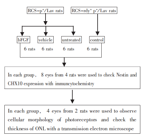

Fig. 1 Animal grouping and experimental procedures

Preparation and Intravitreal Injection of bFGF

bFGF was provided in ampoules at a concentration of 5 μg / 10 μl. Rats were anesthetized. Before intravitreal injection, both eyes were pre-treated with 0.3% levofloxacin hydrochloride eye drops, two times at 5 min intervals, and then 0.1% tetracaine hydrochloride eye drops, also two times at 5 min intervals for conjunctival sac anesthesia. Ten minutes later, under a binocular dissecting microscope, 6 μl bFGF or vehicle was injected into the vitreous body of the right eye, by inserting a 30-gauge needle approximately 1 mm behind the nasal corneal limbus (at a point midway between the ora serrata and the equator of the eyeball, so as to pass through the pars plana of the ciliary epithelium). Care was taken to not injure the iris, lens, ciliary body, or retina. Shortly after the injections, we observed the eyes for signs of hemorrhage or cataract by portable slit-lamp microscope. If either occurred, the animals were eliminated and replaced. After injection, both eyes were treated with 0.3% levofloxacin hydrochloride eye drops 3-4 times per day, at 2 h intervals, for 3 days. Each animal in the bFGF and vehicle groups received a total of three intravitreal injections, as stated above.

Immunohistochemistry

Eight eyes of 4 rats from each of the four groups (total 32 eyes) were enucleated one day after the final intravitreal injection (untreated control animals were prepared at the same biological age) and then fixed by immersion overnight at 4 °C in a mixture of 4% paraformaldehyde and 30% sucrose in 0.1 mol/L phosphate buffer, pH 7.4. Samples were washed three times in phosphate buffer saline (PBS), cryoprotected in PBS plus 30% sucrose, and frozen in CRYOMATRIX embedding medium (CEM) (Thermo Shandon, Pittsburgh, PA). Transverse (or cross-) sections of the retina were cut at 8 μm on a cryotome and thaw-mounted onto Super-Frost glass slides (Fisher Scientific). Sections were air-dried and processed for immunohistochemistry.

Sections were washed three times for 5 min each with PBS, incubated for 15 min in PBS + 0.1% BSA at 37 °C, washed again three times for 5 min each with PBS, and incubated overnight with primary antibody (mouse anti-rat Nestin antibody and rabbit anti-rat Chx10 antibody, Chemicon International, Inc., Temecula, CA, USA) diluted in PBS at 4 °C. Slides were washed three times for 5 min each in PBS and then incubated with fluorescent secondary antibody (FITC-conjugated goat anti-rabbit IgG, Boster Biological Technology, Ltd., Wuhan, China) for 1 h in a humidified chamber at room temperature. Slides were washed four times for 5 min each in cold PBS and mounted on cover slips in 50% glycerol. After that, slides were observed immediately under a fluorescence microscope (ConfoCor2, Zeiss Ltd. Co., Jena, Germany) and all images were captured.

Retinal Histology and Measurement of the Outer Nuclear Layer (ONL) Thickness

Four eyes of 2 rats from each of the four groups (total 16 eyes) were enucleated one day after the final intravitreal injection (untreated control animals were prepared at the same biological age) and fixed in a mixture of 80% ethanol, 40% formalin, and 95% glacial acetic acid (85:10:5 ppv) for 24 h. The eyes were opened by bisecting through the equator, and the posterior eye cups were dehydrated, embedded in paraffin wax, sectioned in the vertical plane close to the optic axis at 4 μm to allow for comparison of all regions of the retina in the superior and inferior hemispheres, mounted onto glass slides, stained with hematoxylin and eosin, and cover-slipped.

The structure of the photoreceptor layer was observed with a light microscope. In each hemisphere, the thickness of the ONL was measured at 480 μm intervals at 4 defined points: at 12:00, 3:00, 6:00, and 9:00 1 mm from the optic nerve head. As measurement of ONL thickness in μm alone can be confounded by tilt of the section plane away from the vertical, besides the expression of ONL thickness in μm, we presented the data in terms of numbers of nuclear lamina, which will correct for any undetected tilting of the section plane. Thus, we expressed the data of ONL thickness in both μm and numbers of nuclear lamina.

Statistical Analysis

SPSS 13.0 software was used to compare the difference among groups. A value was considered significant when P < 0.05.
Results

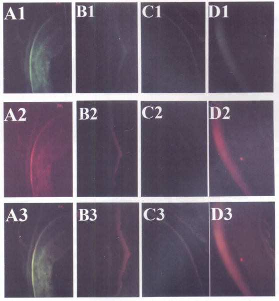
Intravitreal injection of bFGF increased Chx10 and Nestin expression (Fig. 2)


Fig. 2 Typical examples of immunolluorescenee labeling for Chx10 and Nestin in the four experimental groups.
A: bFGF group; B: vehicle group; C: untreated group; D: control group. Intravitreal injection of bfGF in RCS-p+/Lav rats (A) indueed intense labeling for the retinal progenitor cell markers Chx10 (green, Al) and Nestin (red, A2), which were highly eo-loealized (yellowmerged image, A3).Fluorescence intensity for both labels was somewhat less in the untreated, RCS-rdy+p+/Lav rats (D), and muchless in the vehicle-injected RCS rats (B) as well as in the untreated RCS (C). The magnification of all pictures was ×200.

Immunofluorescence coupled to Chx10 and Nestin was present in all retinal layers. The intensity of immunofluorescence was highest in the bFGF treated group, intermediate in the normal control group, and lowest in the vehicle-treated and untreated groups.

    bFGF induced increases in ONL thickness

Under the light microscope, the retinal layers were structurally well defined in all four groups. In the control group, the ONL was about (19.25 ± 2.60) nuclear lamina and (56.23 ± 2.74) μm. The ONL was only about half as thick in the vehicle group [(6.50 ± 1.29) nuclear lamina, (24.86 ± 4.16 μm)] and the untreated group [(6.75 ± 1.71 nuclear lamina, 24.98 ± 2.11 μm)], and no significant difference was observed in ONL thicknesses between vehicle and untreated groups (P > 0.05). Furthermore, while the nuclei themselves appeared normal in these groups, the column of nuclei in their ONL appeared somewhat disorganized. In contrast, in the retina of bFGF-treated RCS, the photoreceptor cell nuclei were small, circular, darkly stained, and regularly arranged, and the ONL was (10.38 ± 2.14) nuclei lamina and (42.52 ± 2.32) μm (Fig. 3). It was significantly thicker in the bFGF group than that in the vehicle group or untreated group (P < 0.01), although still thinner than that of the control group (P < 0.01).

Fig. 3 Retinal histology showed the ONl, thickness of four groups

A: bFGF group; B: vehicle group; C: untreated group; D: control group. The magnification of all the pictures was ×400, HE.

Discussion

Stem cells have the ability to divide (self-replication) for an indefinite time, often throughout the life of the organism. Under the right conditions, or given the right signals, stem cells can give rise (differentiate) to many different cell types that make up the organism. That is, stem cells have the potential to develop into mature cells that have characteristic shapes and specialized functions, such as retinal cells. Typically, stem cells generate an intermediate cell type or types before they achieve their fully differentiated state, and this intermediate cell is called progenitor cells. Progenitor cells in fetal or adult tissues are partly differentiated cells that divide and give rise to differentiated cells. Such cells are usually regarded as “committed” to differentiating along a particular cellular development pathway. Retinitis pigmentosa is mainly caused by mutations that cause damage to photoreceptors, so promoting the process by which stem cells generate progenitor cells, and then in turn photoreceptor cells, should be of benefit for rescuing the affected retina.

In spite of the distinction in regenerative potential between stem cells and progenitor cells, their cell-surface markers (surface proteins that are unique to certain cell types, and capable of detection by antibodies or other detection methods) are largely the same. Nestin, an intermediate filament protein expressed by neural progenitors, is a specific biological marker for progenitor cells, since it is found uniquely in all neural progenitors including those in the retina. Chx10 is a specific marker of nuclei in a subpopulation of retinal progenitor cells and is expressed in divided retinal progenitor cells early in development. The co-expression of Nestin and Chx10 in the retina means the existence of retinal progenitor cells.

bFGF (FGF2) is a protein with 146 amino acids and relative molecular mass 16,500 [16]. In vitro, it increased the proportion of progenitor cells forming clonal primary spheres, and the presence of FGF-2-responsive adult human retinal progenitor cells that divide, express Nestin, produce neurospheres, and differentiate to express markers specific to neuronal, photoreceptor, or glial lineage by 4 times [12-13].

In the present experiment, it showed that intravitreal injection of basic fibroblast growth factor (bFGF) induced increases in immunofluorescence for Chx10 and Nestin, while injection of vehicle alone had little effect. This suggested that bFGF can stimulate endogenous retinal progenitors to self-proliferate and migrate into the central retina. The retina in the control group, at the same age as the other three groups, was well-developed and had no need for retinal progenitor cells to activate and proliferate, so the expression of Chx10 and Nestin was stronger than the vehicle and untreated groups but weaker than the bFGF group. On the other hand, intravitreal injection of bFGF induced an increase in ONL thickness and structural stabilization of photoreceptors, suggesting that bFGF could stimulate endogenous retinal progenitors to differentiate into photoreceptors. Of course, other mechanisms of bFGF may also play a role in maintaining a thick ONL, for example, inhibition of photoreceptor apoptosis [17].


Retina is a well-characterized structure, which is arranged in a stereotypical laminar organization. Neural progenitors with retinal potential are present in the ciliary body epithelium [11, 18], the iris pigment epithelium [
19-20], and the peripheral margin of the postnatal retina [17, 21]. In this study, the site at which bFGF stimulated retinal progenitor cells to reactivate, proliferate, and differentiate was presumed to be in the peripheral retinal margin, since in mammals the iris pigment epithelium is separated from the vitreous body by aqueous humor and the ciliary body is a non-permissive environment for cell migration and neurogenesis [22].


bFGF has well-documented protective neurotrophic activity in rats with photoreceptor degeneration [23-24]. However, although intravitreal injection of bFGF delays photoreceptor degeneration (as in the present experiment), such treatment does not allow long-term rescue of photoreceptors because of the short half-life of the trophic factor. Also, we have no data indicating a dose-response relationship for multiple injections of bFGF. Therefore, further "rescue" studies using bFGF should focus on efficient approaches for continuous delivery, for example, by the trans-corneal eyedrops system that we are developing.

Acknowledgments

The authors thank Dr. Jianming Hu, Dr. Jie Ji, Dr. Jinxiang Huang, and Dr. Yungang Ding for their technical assistance.


1、Friedman DS, O'Colmain BJ, Munoz B, et al. Eye Diseases Prevalence Research Group. Prevalence of age-related macular degeneration in the United States [J]. Arch Ophthalmol, 2004, 122 (4): 564-572.Friedman DS, O'Colmain BJ, Munoz B, et al. Eye Diseases Prevalence Research Group. Prevalence of age-related macular degeneration in the United States [J]. Arch Ophthalmol, 2004, 122 (4): 564-572.
2、Shintani K, Shechtman DL, Gurwood AS. Review and update: current treatment trends for patients with retinitis pigmentosa [J]. Optometry, 2009, 80 (7): 384-401.Shintani K, Shechtman DL, Gurwood AS. Review and update: current treatment trends for patients with retinitis pigmentosa [J]. Optometry, 2009, 80 (7): 384-401.
3、Sakai T, Kuno N, Takamatsu F, et al. Prolonged protective effect of basic fibroblast growth factor-impregnated nanoparticles in Royal College of Surgeons rats [J]. IOVS, 2007, 48: 3381-3387.Sakai T, Kuno N, Takamatsu F, et al. Prolonged protective effect of basic fibroblast growth factor-impregnated nanoparticles in Royal College of Surgeons rats [J]. IOVS, 2007, 48: 3381-3387.
4、Gregory-Evans K, Chang F, Hodges MD, et al. Ex vivo gene therapy using intravitreal injection of GDNF-secreting mouse embryonic stem cells in a rat model of retinal degeneration [J]. Mol Vis, 2009, 15: 962-973.Gregory-Evans K, Chang F, Hodges MD, et al. Ex vivo gene therapy using intravitreal injection of GDNF-secreting mouse embryonic stem cells in a rat model of retinal degeneration [J]. Mol Vis, 2009, 15: 962-973.
5、Beltran WA, Wen R, Acland GM, et al. Intravitreal injection of ciliary neurotrophic factor (CNTF) causes peripheral remodeling and does not prevent photoreceptor loss in canine RPGR mutant retina [J]. Exp Eye Res, 2007, 84 (4): 753-771.Beltran WA, Wen R, Acland GM, et al. Intravitreal injection of ciliary neurotrophic factor (CNTF) causes peripheral remodeling and does not prevent photoreceptor loss in canine RPGR mutant retina [J]. Exp Eye Res, 2007, 84 (4): 753-771.
6、Acland GM, Aguirre GD, Ray J, et al. Gene therapy restores vision in a canine model of childhood blindness [J]. Nat Genet, 2001, 28 (1): 92-95.Acland GM, Aguirre GD, Ray J, et al. Gene therapy restores vision in a canine model of childhood blindness [J]. Nat Genet, 2001, 28 (1): 92-95.
7、Conley S, Nour M, Fliesler SJ, et al. Late-onset cone photoreceptor degeneration induced by R172W mutation in Rds and partial rescue by gene supplementation [J]. IOVS, 2007, 48 (12): 5397-5407.Conley S, Nour M, Fliesler SJ, et al. Late-onset cone photoreceptor degeneration induced by R172W mutation in Rds and partial rescue by gene supplementation [J]. IOVS, 2007, 48 (12): 5397-5407.
8、Bok D. Gene therapy of retinal dystrophies: achievements, challenges and prospects [J]. Novartis Found Symp, 2004, 255: 4-12.Bok D. Gene therapy of retinal dystrophies: achievements, challenges and prospects [J]. Novartis Found Symp, 2004, 255: 4-12.
9、Lund RD, Kwan AS, Keegan DJ, et al. Cell transplantation as a treatment for retinal disease [J]. Prog Retin Eye Res, 2001, 20 (4): 415-449.Lund RD, Kwan AS, Keegan DJ, et al. Cell transplantation as a treatment for retinal disease [J]. Prog Retin Eye Res, 2001, 20 (4): 415-449.
10、Lund R. Cell-based therapies to limit photoreceptor degeneration [J]. Arch Soc Esp Oftalmol, 2008, 83 (8): 457-464.Lund R. Cell-based therapies to limit photoreceptor degeneration [J]. Arch Soc Esp Oftalmol, 2008, 83 (8): 457-464.
11、Tropepe V, Coles BL, Chiasson BJ, et al. Retinal stem cells in the adult mammalian eye. Science, 2000, 287 (5460): 2032-2036.Tropepe V, Coles BL, Chiasson BJ, et al. Retinal stem cells in the adult mammalian eye. Science, 2000, 287 (5460): 2032-2036.
12、Gu P, Harwood LJ, Zhang X, et al. Isolation of retinal progenitor and stem cells from the porcine eye [J]. Mol Vis, 2007, 13: 1045-1057.Gu P, Harwood LJ, Zhang X, et al. Isolation of retinal progenitor and stem cells from the porcine eye [J]. Mol Vis, 2007, 13: 1045-1057.
13、Mayer EJ, Carter DA, Ren Y, et al. Neural progenitor cells from postmortem adult human retina [J]. Br J Ophthalmol, 2005, 89 (1): 102-106.Mayer EJ, Carter DA, Ren Y, et al. Neural progenitor cells from postmortem adult human retina [J]. Br J Ophthalmol, 2005, 89 (1): 102-106.
14、Kumar R, Dutt K. Enhanced neurotrophin synthesis and molecular differentiation in non-transformed human retinal progenitor cells cultured in a rotating bioreactor [J]. Tissue Eng, 2006, 12 (1): 141-158.Kumar R, Dutt K. Enhanced neurotrophin synthesis and molecular differentiation in non-transformed human retinal progenitor cells cultured in a rotating bioreactor [J]. Tissue Eng, 2006, 12 (1): 141-158.
15、Coles BL, Angenieux B, Inoue T, et al. Facile isolation and the characterization of human retinal stem cells [J]. Proc Natl Acad Sci U S A, 2004, 101 (4): 15772-15777.Coles BL, Angenieux B, Inoue T, et al. Facile isolation and the characterization of human retinal stem cells [J]. Proc Natl Acad Sci U S A, 2004, 101 (4): 15772-15777.
16、Esch F, Baird A, Ling N, et al. Primary structure of bovine pituitary basic fibroblast growth factor (FGF) and comparison with the amino-terminal sequence of bovine brain acidic FGF [J]. Proc Natl Acad Sci USA, 1985, 82 (19): 6507-6511.Esch F, Baird A, Ling N, et al. Primary structure of bovine pituitary basic fibroblast growth factor (FGF) and comparison with the amino-terminal sequence of bovine brain acidic FGF [J]. Proc Natl Acad Sci USA, 1985, 82 (19): 6507-6511.
17、Zhao X, Das AV, Soto-Leon F, et al. Growth factor-responsive progenitors in the postnatal mammalian retina [J]. Dev Dyn, 2005, 232 (2): 349-358.Zhao X, Das AV, Soto-Leon F, et al. Growth factor-responsive progenitors in the postnatal mammalian retina [J]. Dev Dyn, 2005, 232 (2): 349-358.
18、Ahmad I, Tang L, Pham H. Identification of neural progenitors in the adult mammalian eye [J]. Biochem Biophys Res Commun, 2000, 270 (2): 517-521.Ahmad I, Tang L, Pham H. Identification of neural progenitors in the adult mammalian eye [J]. Biochem Biophys Res Commun, 2000, 270 (2): 517-521.
19、Asami M, Sun G, Yamaguchi M, et al. Multipotent cells from mammalian iris pigment epithelium [J]. Dev Biol, 2007, 304 (7): 433-446.Asami M, Sun G, Yamaguchi M, et al. Multipotent cells from mammalian iris pigment epithelium [J]. Dev Biol, 2007, 304 (7): 433-446.
20、Sun G, Asami M, Ohta H, et al. Retinal stem/progenitor properties of iris pigment epithelial cells [J]. Dev Biol, 2006, 289 (1): 243-252.Sun G, Asami M, Ohta H, et al. Retinal stem/progenitor properties of iris pigment epithelial cells [J]. Dev Biol, 2006, 289 (1): 243-252.
21、Moshiri%20A%2C%20Reh%20TA.%20Persistent%20progenitors%20at%20the%20retinal%20margin%20of%20ptc%2B%2F%E2%88%92%20mice%20%5BJ%5D.%20J%20Neurosci%2C%202004%2C%2024%20(1)%3A%20229-237.Moshiri%20A%2C%20Reh%20TA.%20Persistent%20progenitors%20at%20the%20retinal%20margin%20of%20ptc%2B%2F%E2%88%92%20mice%20%5BJ%5D.%20J%20Neurosci%2C%202004%2C%2024%20(1)%3A%20229-237.
22、Stokes J, Walker BR, Campbell JC, et al. Altered peripheral sensitivity to glucocorticoid in primary open-angle glaucoma [J]. Invest Ophthalmol Vis Sci, 2003, 44 (12): 5163-5167.Stokes J, Walker BR, Campbell JC, et al. Altered peripheral sensitivity to glucocorticoid in primary open-angle glaucoma [J]. Invest Ophthalmol Vis Sci, 2003, 44 (12): 5163-5167.
23、Xu Yan, Chen Zuji, Song Jiezhen. Comparative study of the rabbit high intraocular pressure model induced by compound carbomer and other rabbit high intraocular pressure models [J]. Chinese Journal of Ophthalmology, 2002, 38 (3): 172-175.Xu Yan, Chen Zuji, Song Jiezhen. Comparative study of the rabbit high intraocular pressure model induced by compound carbomer and other rabbit high intraocular pressure models [J]. Chinese Journal of Ophthalmology, 2002, 38 (3): 172-175.
24、Otte C, Wüst S, Zhao S, et al. Glucocorticoid receptor gene and depression in patients with coronary heart disease: the Heart and Soul Study - 2009 Curt Richter Award Winner [J]. Psychoneuroendocrinology, 2009, 34 (10): 1574-1581.Otte C, Wüst S, Zhao S, et al. Glucocorticoid receptor gene and depression in patients with coronary heart disease: the Heart and Soul Study - 2009 Curt Richter Award Winner [J]. Psychoneuroendocrinology, 2009, 34 (10): 1574-1581.
25、Nehmé A, Lobenhofer EK, Stamer WD, et al. Glucocorticoids with different chemical structures but similar glucocorticoid receptor potency regulate subsets of common and unique genes in human trabecular meshwork cells [J]. BMC Med Genomics, 2009, 2: 58.Nehmé A, Lobenhofer EK, Stamer WD, et al. Glucocorticoids with different chemical structures but similar glucocorticoid receptor potency regulate subsets of common and unique genes in human trabecular meshwork cells [J]. BMC Med Genomics, 2009, 2: 58.
26、Zhang X, Ognibene CM, Clark AF, Yorio T. Dexamethasone inhibition of trabecular meshwork cell phagocytosis and its modulation by glucocorticoid receptor beta [J]. Exp Eye Res, 2007, 84 (2): 275-284.Zhang X, Ognibene CM, Clark AF, Yorio T. Dexamethasone inhibition of trabecular meshwork cell phagocytosis and its modulation by glucocorticoid receptor beta [J]. Exp Eye Res, 2007, 84 (2): 275-284.
27、Hermande MR, Eenk EJ, Weinstein B. Glucocorticoid target cells in human outflow pathway [J]. Invest Ophthalmol Vis Sci, 1983, 24: 1612-1616.Hermande MR, Eenk EJ, Weinstein B. Glucocorticoid target cells in human outflow pathway [J]. Invest Ophthalmol Vis Sci, 1983, 24: 1612-1616.
28、Potau JM, Canals M, Costa J, et al. Ultrastructural characteristics of glaucomatous trabecular meshwork [J]. Arch Soc Esp Oftalmol, 2000, 75 (1): 21-28.Potau JM, Canals M, Costa J, et al. Ultrastructural characteristics of glaucomatous trabecular meshwork [J]. Arch Soc Esp Oftalmol, 2000, 75 (1): 21-28.
29、Johnson D, Gottanka J, Flügel C, et al. Ultrastructural changes in the trabecular meshwork of human eyes treated with corticosteroids [J]. Arch Ophthalmol, 1997, 115 (3): 375-383.Johnson D, Gottanka J, Flügel C, et al. Ultrastructural changes in the trabecular meshwork of human eyes treated with corticosteroids [J]. Arch Ophthalmol, 1997, 115 (3): 375-383.
30、Steely HT, Browder SL, Julian MB, et al. The effects of Dexamethasone on fibronectin expression in cultured human trabecular meshwork cells [J]. Invest Ophthalmol Vis Sci, 1992, 33: 2242-2250.Steely HT, Browder SL, Julian MB, et al. The effects of Dexamethasone on fibronectin expression in cultured human trabecular meshwork cells [J]. Invest Ophthalmol Vis Sci, 1992, 33: 2242-2250.
31、Diekerson JE, Steely HT, English-Wright SL, et al. The effects of dexamethasone on integrin and laminin expression in cultured human trabecular meshwork cells [J]. Exp Eye Res, 1998, 66: 731-738.Diekerson JE, Steely HT, English-Wright SL, et al. The effects of dexamethasone on integrin and laminin expression in cultured human trabecular meshwork cells [J]. Exp Eye Res, 1998, 66: 731-738.
32、Zhuo YH, Huang YL, Wei YT, et al. Glucocorticoids upregulate transepithelial electrical resistance and expression of tight junction-related protein in human trabecular meshwork cells [J]. Chin Med J (Engl), 2005, 118 (20): 1742-1745.Zhuo YH, Huang YL, Wei YT, et al. Glucocorticoids upregulate transepithelial electrical resistance and expression of tight junction-related protein in human trabecular meshwork cells [J]. Chin Med J (Engl), 2005, 118 (20): 1742-1745.
33、Abdouh M, Bernier G. In vivo reactivation of a quiescent cell population located in the ocular ciliary body of adult mammals [J]. Exp Eye Res, 2006, 83 (1): 153-164.Abdouh M, Bernier G. In vivo reactivation of a quiescent cell population located in the ocular ciliary body of adult mammals [J]. Exp Eye Res, 2006, 83 (1): 153-164.
34、Sakai T, Kuno N, Takamatsu F, et al. Prolonged protective effect of basic fibroblast growth factor-impregnated nanoparticles in Royal College of Surgeons rats [J]. IOVS, 2007, 48 (1): 3381-3387.Sakai T, Kuno N, Takamatsu F, et al. Prolonged protective effect of basic fibroblast growth factor-impregnated nanoparticles in Royal College of Surgeons rats [J]. IOVS, 2007, 48 (1): 3381-3387.
35、O'Driscoll C, O'Connor J, O'Brien CJ, et al. Basic fibroblast growth factor-induced protection from light damage in the mouse retina in vivo [J]. J Neurochem, 2008, 105 (2): 524-536.O'Driscoll C, O'Connor J, O'Brien CJ, et al. Basic fibroblast growth factor-induced protection from light damage in the mouse retina in vivo [J]. J Neurochem, 2008, 105 (2): 524-536.
1、Supported by Guangdong Science and Technology Plan Grants#2008B030301300()
上一篇
下一篇
其他期刊
  • 眼科学报

    主管:中华人民共和国教育部
    主办:中山大学
    承办:中山大学中山眼科中心
    主编:林浩添
    主管:中华人民共和国教育部
    主办:中山大学
    浏览
  • Eye Science

    主管:中华人民共和国教育部
    主办:中山大学
    承办:中山大学中山眼科中心
    主编:林浩添
    主管:中华人民共和国教育部
    主办:中山大学
    浏览
推荐阅读
出版者信息
目录