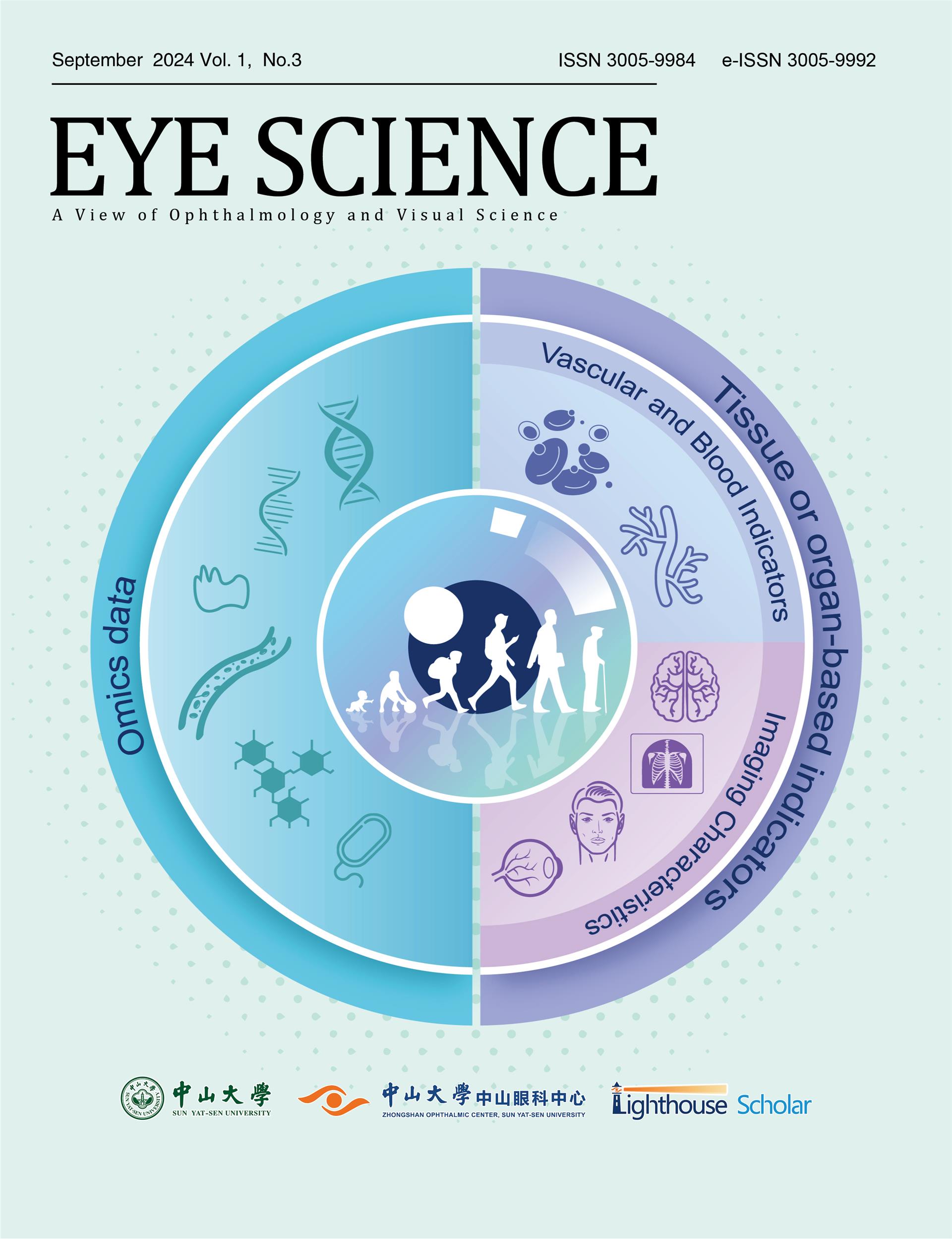
2024年9月 第1卷 第3期

主管:中华人民共和国教育部
主办:中山大学
承办:中山大学中山眼科中心
主编:Haotian Lin, MD, PhD
Commentary

Orthokeratology and the choroid

Orthokeratology and the choroid

:1-5
 
The choroid is a multifunctional, highly vascular, and dynamic tissue which contributes to ocular homeostasis and the regulation of eye growth in both animals and humans. Although challenging to reliably measure, recent advances in ocular imaging (particularly optical coherence tomography) has expanded the current understanding of the role of the choroid in ageing and refractive error development during childhood. This commentary considers recent advances in the field, particularly the impact of orthokeratology on choroidal thickness and contour in myopic children, and the potential use of choroidal metrics as a biomarker for future eye growth.
The choroid is a multifunctional, highly vascular, and dynamic tissue which contributes to ocular homeostasis and the regulation of eye growth in both animals and humans. Although challenging to reliably measure, recent advances in ocular imaging (particularly optical coherence tomography) has expanded the current understanding of the role of the choroid in ageing and refractive error development during childhood. This commentary considers recent advances in the field, particularly the impact of orthokeratology on choroidal thickness and contour in myopic children, and the potential use of choroidal metrics as a biomarker for future eye growth.
Original Article

Machine learning methods for biological age estimation

Machine learning methods for biological age estimation

:176-189
 
Age stands as a primary risk factor for diseases and disabilities among the elderly. To effectively assess the underlying aging processes, accurate measures of biological age and rates of aging across multiple levels of aging features are essential. Biological age derives from physiological assessments of systems and organs. It has emerged as a superior predictor of age-related diseases and mortality compared to chronological age. Recent advancements in machine learning have catalyzed the development of sophisticated models capable of quantitatively characterizing biological aging with different types of data. This review explores the machine learning models in advancing our understanding of biological aging, highlighting the potential of these innovative approaches to facilitate aging research and personalized healthcare strategies.
Age stands as a primary risk factor for diseases and disabilities among the elderly. To effectively assess the underlying aging processes, accurate measures of biological age and rates of aging across multiple levels of aging features are essential. Biological age derives from physiological assessments of systems and organs. It has emerged as a superior predictor of age-related diseases and mortality compared to chronological age. Recent advancements in machine learning have catalyzed the development of sophisticated models capable of quantitatively characterizing biological aging with different types of data. This review explores the machine learning models in advancing our understanding of biological aging, highlighting the potential of these innovative approaches to facilitate aging research and personalized healthcare strategies.

Interaction of ductal obstruction and glandular dropout in the pathogenesis of meibomian gland dysfunction

Interaction of ductal obstruction and glandular dropout in the pathogenesis of meibomian gland dysfunction

:190-205
 
Meibomian gland dysfunction (MGD) manifests through two main clinical presentations, characterized by the meibomian gland (MG) ductal obstruction or acinar dropout. While previous research has predominantly associated MGD pathogenesis with hyperkeratinization-related MG ductal obstruction and subsequent acinar atrophy, recent cases have shown significant functional acinar loss in the absence of apparent ductal keratinization or blockage. The deterioration of either MG obstruction or dropout exacerbates the condition of the other, suggesting an independent yet interconnected relationship that perpetuates the vicious cycle of MGD. Understanding the distinct pathological features of MG obstruction and dropout is crucial for delineating their etiology and identifying targeted therapeutic strategies. This review explores the nuanced interrelations of MG obstruction and dropout, elucidating potential pathological mechanisms to establish a foundation for early MGD diagnosis and intervention.
Review Article

Application and performance of artificial intelligence in screening retinopathy of prematurity from 2018 to 2024: a meta-analysis and systematic review

Application and performance of artificial intelligence in screening retinopathy of prematurity from 2018 to 2024: a meta-analysis and systematic review

:206-223
 
Purpose: Artificial intelligence (AI) significantly enhances the screening and diagnostic processes for retinopathy of prematurity (ROP). In this article,we focused on the application and performance of AI in detecting ROP and distinguishing plus disease (PLUS) in ROP. Methods: We searched PubMed, Embase, Medline, Web of Science, and Ovid for studies published from January 2018 to July 2024. Studies evaluating the diagnostic performance of AI with expert ophthalmologists’judgment as a reference standard were included. The risk of bias was assessed using the QUADAS-2 tool and QUADAS-AI tool.Statistical analysis included data pooling, forest plot construction, heterogeneity testing, and meta-regression. Results: Fourteen of the 186 studies were included.The pooled sensitivity, specificity and the area under the curve (AUC) of the AI diagnosing ROP were 0.95 (95% CI 0.93-0.96), 0.97 (95% CI 0.94-0.98) and 0.97 (95% CI 0.95-0.98), respectively.The pooled sensitivity, specificity and the AUC of the AI distinguishing PLUS were 0.92 (95% CI 0.80-0.97),0.95 (95% CI 0.91-0.97) and 0.98 (95% CI 0.96-0.99), respectively.Cochran’s Q test (< 0.01) andHiggins I heterogeneity index revealed considerable heterogeneity. The country of study, number of centers, data source and the number of doctors were responsible for the heterogeneity. For ROP diagnosing, researches conducted in China using private data in single center with less than 3 doctors showed higher sensitivity and specificity. For PLUS distinguishing, researches in multiple centers with less than 3 doctors showed higher sensitivity. Conclusions: This study revealed the powerful role of AI in diagnosing ROP and distinguishing PLUS. However, significant heterogeneity was noted among all included studies, indicating challenges in the application of AI for ROP diagnosis in real-world settings. More studies are needed to address these disparities, aiming to fully harness AI’s potential in augmenting medical care for ROP.
Purpose: Artificial intelligence (AI) significantly enhances the screening and diagnostic processes for retinopathy of prematurity (ROP). In this article,we focused on the application and performance of AI in detecting ROP and distinguishing plus disease (PLUS) in ROP. Methods: We searched PubMed, Embase, Medline, Web of Science, and Ovid for studies published from January 2018 to July 2024. Studies evaluating the diagnostic performance of AI with expert ophthalmologists’judgment as a reference standard were included. The risk of bias was assessed using the QUADAS-2 tool and QUADAS-AI tool.Statistical analysis included data pooling, forest plot construction, heterogeneity testing, and meta-regression. Results: Fourteen of the 186 studieswere included.The pooled sensitivity, specificity and the area under the curve (AUC) of the AI diagnosing ROP were 0.95 (95% CI 0.93-0.96), 0.97 (95% CI 0.94-0.98) and 0.97 (95% CI 0.95-0.98), respectively.The pooled sensitivity, specificity and the AUC of the AI distinguishing PLUS were 0.92 (95% CI 0.80-0.97),0.95 (95% CI 0.91-0.97) and 0.98 (95% CI 0.96-0.99), respectively.Cochran’s Q test (< 0.01) andHiggins I heterogeneity index revealed considerable heterogeneity. The country of study, number of centers, data source and the number of doctors were responsible for the heterogeneity. For ROP diagnosing, researches conducted in China using private data in single center with less than 3 doctors showed higher sensitivity and specificity. For PLUS distinguishing, researches in multiple centers with less than 3 doctors showed higher sensitivity. Conclusions: This study revealed the powerful role of AI in diagnosing ROP and distinguishing PLUS. However, significant heterogeneity was noted among all included studies, indicating challenges in the application of AI for ROP diagnosis in real-world settings. More studies are needed to address these disparities, aiming to fully harness AI’s potential in augmenting medical care for ROP.

Trends and hotspots concerning lupus retinopathy from 2003 to 2022: a bibliometric analysis and knowledge graph study

Trends and hotspots concerning lupus retinopathy from 2003 to 2022: a bibliometric analysis and knowledge graph study

:224-236
 
Purpose: To explore the status of current global research, trends and hotspots in the field of lupus retinopathy (LR). Methods: Publications related to LR from 2003 to 2022 were extracted from the Web of Science Core Collection (WOSCC). Citespace 6.2.R4 software was used to analyze the raw data. Bibliometric parameters such as publication quality, countries, authors, international cooperation, and keywords were taken into account. Results: A total of 315 publications were retrieved. The annual research output has increased significantly since 2010, especially since 2017. Marmor MF, Lee BR, and Melles RB contributed the highest number of articles published on LR. The top three publishing countries were the USA, China, and UK. Stanford University, Hanyang University, and Harvard Medical School were the top three producing institutions in the world for LR research. The top ten commonly used keywords include the following: systemic lupus erythematosus, retinopathy, retinal toxicity, antimalarial, hydroxychloroquine, optical coherence tomography, antiphospholipid syndrome, microvascular, optic neuritis, optical coherence tomography angiography. The keywords "optical coherence tomography angiography" and "vessel density" have exploded in recent years. Conclusion: By analyzing the current body of LR literature, specific global trends and hotspots for LR research were identified, presenting valuable information to track cutting- edge progress and for future cooperation between various authors and institutions.

Progression of visual impairment in a patient harboring OPA1 mutation: a case report and literature review

Progression of visual impairment in a patient harboring OPA1 mutation: a case report and literature review

:237-243
 
Dominant optic atrophy (DOA) is an inherited optic neuropathy and more than 75% of DOA patients harbor pathogenic mutations in OPA1. We reported a 39-year-old female harboring c.2119G>T mutation of OPA1 and manifested progressive visual impairment after hydroxychloroquine (HCQ) therapy. The patient’s visual impairment remained stable for 10 years until she began to take HCQ 13 months ago. She complained about progressively decreased vision in both eyes. Bilateral pale temporal optic disc was similar with that of 11 years ago. Optical coherence tomography showed bilateral moderate retinal nerve fiber layer thinning other than the nasal quadrant and general thinning of the inner retina in the macular. Microcystic macular edema was noted in nasal macular in both eyes. Visual field testing showed paracentral scotoma and microperimetry showed decrease sensitivity in the macular in both eyes. After the patient stopped taking HCQ, her functional tests including visual acuity, field testing and microperimetry testing was stable compared with those of 2 years ago. However, progressive inner macular and RNFL thinning was shown by OCT. OPA1 c.2119 G>T found in this patient was a mutation that had been rarely reported in previous studies. The patient has been followed up for over 10 years and her visual acuity stayed stable for decades long until she took HCQ for 13 months. Her vision decline terminated after she stopped taking HCQ. Although HCQ toxicity is highly related to the duration and daily dose, HCQ may aggravate visual impairment in certain individuals harboring OPA1 mutation. Patients with DOA should avoid using neurotoxic HCQ and other medications that may interfere mitochondrial metabolism.

Dominant optic atrophy (DOA) is an inherited optic neuropathy and more than 75% of DOA patients harbor pathogenic mutations in OPA1. We reported a 39-year-old female harboring c.2119G>T mutation of OPA1 and manifested progressive visual impairment after hydroxychloroquine (HCQ) therapy. The patient’s visual impairment remained stable for 10 years until she began to take HCQ 13 months ago. She complained about progressively decreased vision in both eyes. Bilateral pale temporal optic disc was similar with that of 11 years ago. Optical coherence tomography showed bilateral moderate retinal nerve fiber layer thinning other than the nasal quadrant and general thinning of the inner retina in the macular. Microcystic macular edema was noted in nasal macular in both eyes. Visual field testing showed paracentral scotoma and microperimetry showed decrease sensitivity in the macular in both eyes. After the patient stopped taking HCQ, her functional tests including visual acuity, field testing and microperimetry testing was stable compared with those of 2 years ago. However, progressive inner macular and RNFL thinning was shown by OCT. OPA1 c.2119 G>T found in this patient was a mutation that had been rarely reported in previous studies. The patient has been followed up for over 10 years and her visual acuity stayed stable for decades long until she took HCQ for 13 months. Her vision decline terminated after she stopped taking HCQ. Although HCQ toxicity is highly related to the duration and daily dose, HCQ may aggravate visual impairment in certain individuals harboring OPA1 mutation. Patients with DOA should avoid using neurotoxic HCQ and other medications that may interfere mitochondrial metabolism.

出版者信息